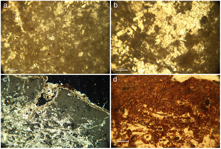
Fig 6. Micrographs of bone thin sections illustrating various histotaphonomic characteristics.
a) Amalgamations of non-Wedl micro-foci of destruction (MFD) in sample PAL011 which appear as dark ‘cloudy’ masses of erosion surrounding the osteons, and stained infiltrations in some Haversian canals; b) Wedl MFD in sample GS19 observed as dark and meandering erosive defects, some of which emanate from cracks in the bottom left quarter of the image; c) Inclusions in sample GS36 which are identified as bright (birefringent) white and golden coloured structures on the external/periosteal surface of the bone (from top left to middle right) and within the micropores and cracks in the mesosteal third of the bone (middle to bottom left) under polarised light; d) Probable iron oxide staining in sample CND025 presents as mosaic-like formations of dark reddish-brown intrusions, distributed thickly along the periosteal band and more sparsely in the mesosteal third of the sample (bottom half of the image). All figures author’s own, published under CC BY license.

