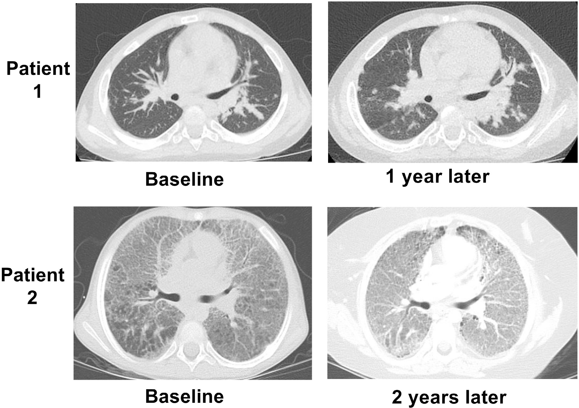
Figure 5.

Variability in imaging progression in patients with SJIA-LD. For patient 1, baseline chest CT demonstrated extensive parenchymal pulmonary opacities with interstitial thickening. Repeat imaging showed an overall similar pattern with development of small cysts suggesting fibrosis despite stopping all anticytokine therapy. Patient 2 at baseline demonstrated extensive bilateral ground-glass opacities and interlobular septal thickening. Follow-up study 2 years later showed progression of findings, including more dependent consolidation despite treatment with multiple biologic and nonbiologic agents. CT, computed tomography; LD, lung disease; SJIA, systemic juvenile idiopathic arthritis.
