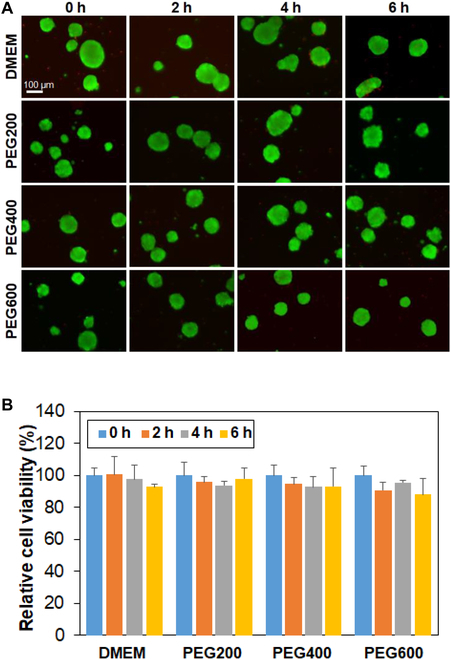
Fig. 3.

Cytotoxicity assay of PEGs for SCSs. (A) Live/dead images of SCSs in DMEM containing PEGs. The SCSs were suspended in the solution and incubated at 37 °C for 0 h, 2 h, 4 h, and 6 h. The scale bar is 100 μm. (B) Cell viability assayed using the CCK-8 kit. The relative cell viability was compared with 0 h (100%).
