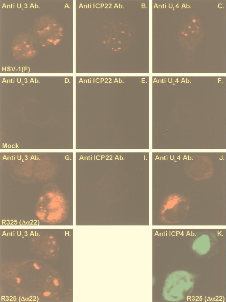
FIG. 3.
Localization of UL3 and UL4 proteins and of ICP22 in infected cells. Rabbit skin cells were mock infected or exposed to 10 PFU of HSV-1(F) or R325 per cell and processed as described in the legend to Fig. 1. Anti-mouse immunoglobulin conjugated to FITC (Sigma) was used to visualize the mouse monoclonal antibody to ICP4 (K). Ab., antibody.

