



Abstract

Lactononadecapeptide (LNDP; NIPPLTQTPVVVPPFLQPE), a casein-derived peptide comprising 19 residues, is known for its capacity to enhance cognitive function. This study aimed to explore the transepithelial transport and stability of LNDP. Results showed that LNDP retained over 90% stability after 2 h of treatment with gastrointestinal enzymes. The stability of LNDP on Caco-2 cell monolayers ranged from 93.4% ± 0.9% to 101.1% ± 1.2% over a period of 15–60 min, with no significant differences at each time point. The permeability of LNDP across an artificial lipid membrane was very low with the effective permeability of 3.6 × 10⁻¹¹ cm/s. The Caco-2 assay demonstrated that LNDP could traverse the intestinal epithelium, with an apparent permeability of 1.22 × 10⁻⁶ cm/s. Its transport was significantly inhibited to 67.9% ± 5.0% of the control by Gly-Pro, a competitor of peptide transporter 1 (PEPT1). Furthermore, PEPT1 knockdown using siRNA significantly inhibited LNDP transport by 77.6% ± 1.9% in Caco-2 cell monolayers. The LNDP uptake in PEPT1-expressing HEK293 cells was significantly higher (54.5% ± 14.6%) than that in mock cells. These findings suggest that PEPT1 plays a crucial role in LNDP transport, and LNDP exhibits good resistance to gastrointestinal enzymes.
Keywords: peptide, beta-casein, lactononadecapeptide (LNDP), Caco-2 cells, intestinal transport, peptide transporter 1 (PEPT1)
Introduction
Several bioactive peptides have been identified in various food proteins. Notably, milk-derived peptides exhibit multiple physiological activities, including hypotensive,1,2 immunomodulatory,3,4 and cognitive function improvement effects.5,6 These active sequences range in size from 2 to 20 amino acid residues.
Orally consumed peptides, whether in food or supplements, must reach their target sites in an active form to be functional. Typically, peptide absorption occurs primarily through the epithelium of the small intestine after gastrointestinal digestion. It is essential to verify whether peptides are degraded by digestive enzymes like pepsin, pancreatin, or brush border peptidases. However, some bioactive peptides, especially those containing proline, show resistance to proteolysis.7−9 Key factors influencing intestinal permeation include resistance to gastrointestinal enzymes and permeability through the intestinal epithelium.7,10 Factors determining intact absorption of peptides from the intestinal epithelium include molecular weight, hydrophobicity, charge, and aggregation tendency.11,12
There are three primary pathways for peptide transport across the intestinal cell monolayer membrane: passive extracellular diffusion through tight junctions (TJs), transcytosis via vesicles, and active transport via the PEPT1 transporter.11,13−15 The transepithelial transport pathway varies with the peptide.
Peptide permeability studies typically use two assays: the parallel artificial membrane permeability assay (PAMPA) and cell monolayer assays like the Caco-2 assay. Caco-2 cells, a human intestinal carcinoma cell line, have long been used to simulate the structure and function of the human intestinal epithelium in vitro.(16−18) They are instrumental in testing drug absorption mechanisms through the intestinal epithelium. Notably, a strong correlation has been established between passive drug transport through the Caco-2 cell monolayer and in vivo absorption.19
Previously, we discovered that Asn-Ile-Pro-Pro-Leu-Thr-Gln-Thr-Pro-Val-Val-Val-Pro-Pro-Phe-Leu-Gln-Pro-Glu (NIPPLTQTPVVVPPFLQPE, lactononadecapeptide; LNDP), a 19-residue peptide derived from milk beta-casein, enhances cognitive function.20,21 Various human studies have validated LNDP’s cognitive benefits in healthy individuals. For instance, an 8-week supplementation of healthy middle-aged adults (50–70 years old) with a Lactobacillus helveticus-fermented milk drink containing LNDP (2.4 mg/day) improved attention and delayed memory.22 Additionally, our research demonstrated that a 12-week supplementation with LNDP tablets (2.1 mg/day) enhanced cognitive functions, such as attention, concentration, and information processing, in healthy middle-aged and senior adults (60–79 years old).23 Furthermore, a single dose of LNDP tablets (2.4 mg) was found to boost memory, concentration, and attention in healthy adult males (30–59 years old) under cognitive stress.24 The mechanism behind LNDP-induced enhancement of cognitive function involves increasing acetylcholine release and brain-derived neurotrophic factor gene expression in hippocampal neurons.25,26
However, the intestinal transport mechanism of LNDP remains unexplored. Therefore, we investigated both the transport pathway of LNDP through the intestine and its resistance to gastrointestinal enzymes.
Materials and Methods
Chemicals
Antipyrine (99% purity), Eagle’s Minimal Essential Medium (EMEM), Penicillin-Streptomycin (PS), Acetonitrile (ACN), and Perchloric Acid were sourced from Wako Pure Chemical Industries, Ltd. (Osaka, Japan). Hanks Balanced Salt Solution (HBSS) with calcium and magnesium, 4-(2-Hydroxyethyl)-1-piperazineethanesulfonic acid (HEPES), and Lucifer Yellow (LY) were acquired from Thermo Fisher Scientific (Waltham, MA, USA). Fetal Bovine Serum (FBS) was supplied by Biowest (Nuaillé, France). Nonessential Amino Acids (NEAA) were obtained from Gibco (Burlington, ON, Canada). LNDP with 95% purity was synthesized by Funakoshi Co., Ltd. (Tokyo, Japan). The Gly-Pro peptide (GP) (99% purity) was procured from BACHEM (Bubendorf, Switzerland). Wortmannin (98% purity) and Cytochalasin D (95% purity) were purchased from TOCRIS (Bristol, UK). Triton X-100 was obtained from Sigma-Aldrich Co. LLC (St. Louis, MO, USA). Verapamil (98% purity) and Theophylline (98% purity) were acquired from Tokyo Chemical Industry Co., Ltd. (Tokyo, Japan).
LNDP Quantification
The quantification of LNDP via LC-MS/MS analysis was conducted following a previously described method, with some modifications.22 Specifically, the calibration curve’s concentration range was adjusted to enhance sensitivity at lower concentrations. Standard peptides were prepared in 20% ACN at concentrations of 10, 20, 40, 80, 160, 320, 640, 1,280, 2,560, and 5,120 pM. Internal standard peptides were dissolved in 20% ACN at a concentration of 4 nM. For analysis, 90 μL of standard peptides were mixed with 10 μL of analytical internal standard (IS) for LC-MS/MS analysis. This process was carried out using a SCIEX Triple Quad 6500+ system (Framingham, MA, USA).
In Vitro Gastrointestinal Digestion
LNDP was dissolved to a concentration of 10 μM. It underwent digestion with pepsin (enzyme-to-substrate ratio = 1:25) at 37 °C for 2 h (pH 2.0). Similarly, digestion with pancreatin (enzyme-to-substrate ratio = 1:25) was carried out at 37 °C for 2 h (pH 7.2). To halt enzyme activity, the mixture was heat-denatured for 10 min postdigestion. Subsequently, analysis of LNDP was conducted. The digested products were also detected and analyzed.27
PAMPA Experiment
PAMPA was conducted using 96-well filter plates (Millipore, Billerica, MA, USA). Both the donor and acceptor plates were filled with 350 μL of 1% BSA in PBS. The samples were washed twice with Milli-Q water. Subsequently, 5 μL of 20 mg/mL lipid solution in dodecane and 200 μL of PBS were added to the acceptor plate, and 300 μL of the sample in PBS was added to the donor plates. The plates were then incubated at room temperature (25 °C) for 2 h. Postincubation, the LNDP concentration was measured using LC-MS/MS. The assay was carried out in triplicate. Following this, both donor and acceptor solutions were collected, and the effective permeability (Pe) was calculated.
Cell Culture
Caco-2 cells, obtained from the Riken Cell Bank, were cultured in EMEM supplemented with 10% FBS, 1% NEAA, and 1% PS. The cells were maintained in a humidified incubator at 37 °C and 5% CO2. For transport experiments, Caco-2 cells were seeded at a density of 1 × 10⁵ cells/well in falcon permeable support for 24-well plate and allowed to differentiate for 21 days.28 The integrity of the cell layer was verified by measuring Transepithelial Electrical Resistance (TEER) using a Millicell-ERS (Merck Millipore Corporation, Darmstadt, Germany). Only Caco-2 monolayers exhibiting TEER values greater than 250 Ω × cm2 were utilized in the experiments.
Transepithelial Transport Studies
Prior to transport experiments, Caco-2 cells were rinsed with transport buffer (TB, HBSS supplemented with 10 mM HEPES). For the experiment, 10 μM LNDP, dissolved in 0.2 mL of TB, was added to the apical side of the cell monolayers, while 0.8 mL of TB was added to the basal side. The cells were then incubated at 37 °C for durations of 15, 30, or 60 min. Upon completion of the incubation period, samples from both the apical and basal solutions were collected. The permeability of LNDP was calculated by measuring its concentration using LC-MS/MS analysis.
For the inhibition experiments, three inhibitors were used: GP (a PEPT1 substrate), wortmannin (a transcytosis inhibitor), and cytochalasin D (a tight junction disruptor). GP was prepared as a 10 mM solution in TB, while wortmannin (0.5 mM) and cytochalasin D (0.5 mg/mL) were dissolved in TB containing 0.05% DMSO. Each inhibitor (GP, wortmannin, or cytochalasin D) was preincubated for 30 min, followed by a 60 min incubation with 10 μM LNDP on the apical side and TB on the basal side. After the incubation, both apical and basal solutions were collected for analysis. The apparent permeability (Papp) of each setup was then calculated.
Knockdown of PEPT1 in Caco-2 Cells
To inhibit PEPT1 expression in cell monolayers, siRNA targeting PEPT1 was utilized. The sequence for the PEPT1-targeting siRNA was 5′-GCAUCGGAGUAAGGCAUUUTT-3′ (sense) and 5′-AAAUGCCUUACUCCGAUGCCT-3′ (antisense). For control purposes, a group treated with scrambled siRNA, having a disordered sequence, was used. The scrambled siRNA had the following sequences: 5′-GCGCGCUUUGUAGGAUUCGDTDT-3′ (sense) and 3′-DTDTCGCGCGAAACAUCCUAAGC-5′ (antisense).
The siRNA inhibition experiments followed the protocol outlined in a previous study.28 Briefly, 1 day before the transport assay, the cell monolayers were washed twice with OPTI-MEM medium. Then, 500 μL of OPTI-MEM containing 1 μL of Lipofectamine 3000 and 80 nM siRNA was added to the apical side of the cells. After 6 h of transfection, the transfection medium was replaced with growth medium, and the cells were incubated for an additional 24 h. The monolayers were then rinsed three times and preincubated with HBSS for 30 min. Following this, transport assays commenced with an incubation in 10 μM LNDP, and samples from both the apical and basal sides were collected after 60 min of incubation.
Uptake Experiment
To determine if LNDP is a substrate for PEPT1, we conducted uptake experiments using two types of cells: human PEPT1 (hPEPT1) cells and mock cells. hPEPT1 cells are HEK293 cells transfected with a vector containing human PEPT1 cDNA, whereas mock cells are HEK293 cells transfected with an empty vector. The uptake experiment was performed in special, ready-to-use cell plates, procured from Genomembrane Co., Ltd. (Kanagawa, Japan), enabling us to perform assays directly on the received plates. The uptake experiments followed the protocol accompanying the plate products. Initially, the cells were washed twice with the provided buffer solution and preincubated for 5 min. After removing the buffer solution, 10 μM LNDP, dissolved in the buffer, was added to initiate the experiment. The cells were incubated at 37 °C for 5 min following the addition of the LNDP solution. Postincubation, the cells were washed three times with a cold incubation solution that did not contain LNDP. The cells were then lysed in 200 μL of 0.1% Triton X solution. The lysate was centrifuged at 10,000 rpm for 5 min, and the resultant supernatant was collected for subsequent LC-MS/MS analysis.
Data Analysis
Values are presented as means ± SE. For statistical comparison between two groups, an unpaired Student’s t-test was utilized, while Dunnett’s test was employed for analyzing three or more groups. *P < 0.05 was considered statistically significant. All statistical analyses were conducted using SPSS software (version 24.0; IBM Inc., Chicago, IL., USA).
Pe (cm/s) was calculated using the following formula:
 |
where A is the active surface area (0.3 cm2), Vd and Va are the well volumes of the donor and acceptor plates (0.3 and 0.2 cm2, respectively), t is the incubation time (7,200 s), and Cd(t) and Ca(t) are the concentrations of LNDP in the donor and acceptor plates at time t, respectively.
Papp (cm/s) was calculated as described in previous reports:28,29
 |
where dQ/dt is the permeability rate (μmol/s) on the basal side; A is the monolayer surface area (cm2); C0 is the initial concentration on the apical side (μM).
Results and Discussion
Stability of LNDP during in Vitro Digestion
The recovery rates of LNDP following exposure to pepsin and pancreatin for 2 h were 93.2% ± 0.7% and 99.2% ± 1.1%, respectively. This indicates that LNDP is not readily degraded by digestive tract enzymes (Figure 1a). Many orally active peptides reach the bloodstream through normal digestive processes, and it is known that hydrophobic peptides are resistant to degradation. The stability of LNDP could be attributed to its hydrophobic nature. According to the “Keil rule,” trypsin in pancreatin cleaves arginine or lysine but not proline.30−32 Moreover, it is reported that peptides rich in proline and glutamate are gastrointestinal resistant.33 Therefore, the presence of six proline and one glutamate residues in LNDP’s sequence may contribute to its resistance to trypsin and absorption in its intact form. Additionally, to maintain their physiological activities, peptides must exhibit resistance to brush-border peptidases. Hence, sensitivity to intracellular peptidases is a crucial factor in determining the transport of oligopeptides.34 The stability of LNDP in Caco-2 cells, ranged from 93.4% ± 0.9% to 101.1% ± 1.2% over a period of 15–60 min, with no significant differences at each time point, indicating minimal degradation by brush border membrane peptidases (Figure 1b).
Figure 1.

Stability of LNDP (10 μM). All data are presented as means ± SE (n = 3). (a) Recovery rate of LNDP following exposure to digestive tract gastrointestinal enzymes. (b) Recovery rate of LNDP on the apical side of Caco-2 cell monolayers within 60 min.
Transepithelial Transport of LNDP
PAMPA experiments were conducted to assess whether LNDP permeates by passive diffusion. The results, presented in Table 1, show that the Pe for LNDP was 3.6 × 10⁻¹¹ cm/s. For comparison, the Pe of verapamil, a highly permeable sample, was 2.0 × 10⁻⁵ cm/s, while the low permeability samples theophylline and antipyrine had Pe values of 1.7 × 10⁻⁸ and 7.3 × 10⁻⁷ cm/s, respectively. This indicates that LNDP exhibits very low artificial lipid membrane permeability and does not permeate via passive diffusion. It has been reported that peptides with large molecular weights are less likely to pass through cell membranes.35 It is reasonable to assume that LNDP is not passively transported due to its large molecular weight of 2086.4. In investigating LNDP permeability through Caco-2 cell monolayers, LNDP was collected from both the apical and basal side after 15, 30, and 60 min of incubation. The transport percentage of LNDP gradually increased to 0.05% ± 0.03%, 0.39% ± 0.17%, and 0.71% ± 0.17% at 15, 30, and 60 min, respectively (Figure 2a), suggesting that LNDP can be transported across the intestinal mucosa from the lumen to the plasma. Figure 2b reveals no significant difference in LNDP transport between treatments with Cytochalasin D and wortmannin (P > 0.05), indicating that LNDP is not passively transported by paracellular diffusion or transcytosis through Caco-2 cell monolayers. However, LNDP transport was significantly reduced (P < 0.05) in the presence of GP, limiting the transport rate to 67.9% ± 5.0%, suggesting that LNDP transport is mediated by PEPT1.
Table 1. Permeability of an Artificial Lipid Membrane Using PAMPAa.
| Compounds | Average (cm/s) | SE (cm/s) | Reported Average (cm/s)49 |
|---|---|---|---|
| Verapamil | 2.0 × 10⁻⁵ | 1.2 × 10⁻⁶ | 1.4 × 10⁻⁵ |
| Theophylline | 1.7 × 10⁻⁸ | 1.0 × 10⁻⁹ | 4.3 × 10⁻⁸ |
| Antipyrine | 7.3 × 10⁻⁷ | 3.8 × 10⁻⁸ | 8.2 × 10⁻⁷ |
| LNDP | 3.6 × 10⁻¹¹ | 2.7 × 10⁻¹¹ |
The incubation was carried out for 2 h at room temperature. All values represent means ± SE (n = 3).
Figure 2.

Transepithelial transport of LNDP in Caco-2 cells. (a) Impact of incubation time on LNDP transport. Data are presented as means ± SE (n = 3). (b) Effects of GP, Cytochalasin D, and Wortmannin on LNDP transport. Values are means ± SE (*P < 0.05 compared to the control group, n = 18).
The transepithelial transport pathway varies for different peptides. For example, Gln-Ile-Gly-Leu-Phe (QIGLF),36 Arg-Val-Pro-Ser-Leu (RVPSL),37 Val-Leu-Pro-Val-Pro (VLPVP),29 and Trp-Gly-Ala-Pro-Ser-Leu (WGAPSL)38 predominantly utilize TJs for transport. The 17-residue peptide Tyr-Gln-Glu-Pro-Val-Leu-Gly-Pro-Val-Ala-Gly-Pro-Phe-Pro-Ile-Ile-Val (YQEPVLGPVAGPFPIIV) resists digestion by brush border membrane peptidases, and its primary transport pathway is through internalized vesicles via transcytosis in Caco-2 cell monolayers.18 The main transport pathways for the tripeptides Ile-Gln-Trp (IQW), Leu-Lys-Pro (LKP), and Tyr-Pro-Ile (YPI) are likely mediated by PEPT1.28,39
The dipeptide GP is a classical competitive inhibitor of PEPT1 and has a low Km value. PEPT1, an H+-coupled transporter, is primarily located in the gastrointestinal membrane.11 Generally, the PEPT1 pathway facilitates the absorption of dipeptides, tripeptides, and peptide-like drugs, such as the antiviral drug valacyclovir and the antitumor drug ubenimex.40 Amino groups, carboxyl groups, and peptide bonds typically characterize PEPT1 substrates.40 However, even molecules lacking these groups can be substrates for PEPT1, indicating its broad substrate specificity. PEPT1 also plays a role in transporting large peptide prodrug molecules, such as valacyclovir.41 Despite this, it was unclear whether PEPT1 could transport peptides longer than three residues. The significant inhibition of 19-residue LNDP peptide transport by GP suggests that LNDP is absorbed via the PEPT1 pathway. This finding aligns with other research indicating that some peptides with four or more residues are transported via PEPT1; examples include Val-Leu-Pro-Val-Pro-Gln-Lys (VLPVPQK), Val-Leu-Pro-Val-Pro-Gln (VLPVPQ), Ile-Pro-Ala-Val (IPAV), Ile-Pro-Tyr-Trp-Thr-Tyr (IPYWTY), and Val-Pro-Leu-Val-Met (VPLVM).27,42−44 It is evident that PEPT1 can transport a diverse range of substrates, with the spatial arrangement between the Cα atoms of the peptide being a critical factor.40,45 The intestinal absorption of peptides, particularly those containing tertiary amide bonds, is influenced by cis/trans isomerization, as only trans conformational isomers are transported. While normal peptide bonds are predominantly in the trans form due to steric hindrance, proline peptide bonds can adopt both cis and trans conformations. The cis/trans combination of the six proline residues in LNDP potentially enhances its suitability as a substrate for PEPT1.
Effects of PEPT1 siRNA and PEPT1-High Expressing Cells on LNDP Transport
To further investigate whether LNDP transport is mediated by PEPT1, we conducted two experiments. Initially, siRNA was utilized to inhibit PEPT1 expression in Caco-2 cells. LNDP transport was reduced by 77.6% ± 1.9% in these cells compared to a Papp value of 1.22 × 10⁻⁶ cm/s in control cells (P < 0.05, Figure 3). This study was based on previous literature that reported a 54% reduction in PEPT1 protein expression under similar knockdown conditions.28 The decrease in LNDP transport by the PEPT1 siRNA treatment suggested that LNDP may be transported though PEPT1. Subsequently, to corroborate that LNDP is a PEPT1 substrate, we compared LNDP uptake between hPEPT1 and mock HEK293 cells. The hPEPT1 cells exhibited 54.5% ± 14.6% higher uptake of LNDP at 5 min compared to mock cells which exhibited an LNDP uptake of 0.021 ± 0.000 pmol/mg protein (P < 0.05, Figure 4), indicating a potential involvement of PEPT1 in LNDP transport.
Figure 3.
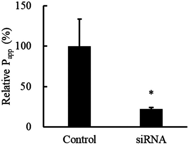
Influence of siRNA targeting PEPT1 on LNDP transport across Caco-2 cell monolayers. Postincubation, LNDP concentration was assessed via LC-MS/MS. Values are means ± SE (*P < 0.05 compared to the control group, n = 12).
Figure 4.

Uptake of LNDP into hPEPT1 cells versus mock HEK293 cells. Cells were incubated at 37 °C for 5 min. LNDP concentration postincubation was determined using LC-MS/MS. All data are means ± SE (*P < 0.05 compared to mock cells, n = 6).
Papp values are employed to assess peptide permeability in Caco-2 cell monolayers. Typically, Papp values for normal peptides range between 10–9 and 10–6 cm/s.27 For example, the Papp values for VLPVP, QIGLF, and RVPSL peptides were recorded as 7.44 × 10⁻⁸, 9.11 × 10⁻⁷, and 6.97 × 10⁻⁶ cm/s, respectively.29,36,37 As depicted in Figure 3, the Papp for LNDP was 1.22 × 10⁻⁶ cm/s, which is higher than those of other peptides, suggesting that LNDP exhibits good permeability.
Numerous prodrug studies have focused on optimizing interactions with PEPT1 by enhancing binding selectivity. It has been established that inclusion of valine or isoleucine often leads to increased cellular permeability.46 For instance, compounds like verapamil or floxuridine have shown increased uptake through binding to valine or isoleucine in PEPT1-high expressing cells.46−48 The presence of valine or isoleucine in LNDP’s amino acid sequence may contribute to its enhanced uptake in cells expressing high levels of PEPT1. However, other mechanisms might also play a role, given that the location of valine or isoleucine in LNDP is internal rather than terminal.
In summary, this study explored the mechanism of LNDP transport through the intestinal tract using Caco-2 cell cultures. It suggests that LNDP is transported through intestinal epithelium in its intact form and into the bloodstream via an active transport pathway mediated by PEPT1 (Figure 5). To the best of our knowledge, this is one of the first reports of relatively long-residue peptides (more than 10 amino acids) being transported through the intestinal tract via PEPT1, warranting further investigation.
Figure 5.

Schematic diagram of the transepithelial transport pathway for the 19-residue peptide LNDP across enterocyte monolayers: Active transport via PEPT1.
Glossary
Abbreviations Used
- ACN
Acetonitrile
- EMEM
Eagle’s Minimal Essential Medium
- FBS
Fetal Bovine Serum
- GP
Gly-Pro
- HBSS
Hanks Balanced Salt Solution
- HEPES
4-(2-Hydroxyethyl)-1-piperazineethanesulfonic Acid
- hPEPT1
Human Peptide Transporter 1
- IPAV
Ile-Pro-Ala-Val
- IPYWTY
Ile-Pro-Tyr-Trp-Thr-Tyr
- IQW
Ile-Gln-Trp
- LKP
Leu-Lys-Pro
- LNDP
Lactononadecapeptide (Asn-Ile-Pro-Pro-Leu-Thr-Gln-Thr-Pro-Val-Val-Val-Pro-Pro-Phe-Leu-Gln-Pro-Glu; NIPPLTQTPVVVPPFLQPE)
- LY
Lucifer Yellow
- NEAA
Nonessential Amino Acids
- PAMPA
Parallel Artificial Membrane Permeability Assay
- Papp
Apparent Permeability Coefficient
- Pe
efficient permeability
- PS
Penicillin-Streptomycin
- PEPT1
Peptide Transporter 1
- QIGLF
Gln-Ile-Gly-Leu-Phe
- RVPSL
Arg-Val-Pro-Ser-Leu
- TB
Transport Buffer
- TEER
Transepithelial Electrical Resistance
- TJs
Tight Junctions
- VPLVM
Val-Pro-Leu-Val-Met
- VLPVP
Val-Leu-Pro-Val-Pro
- VLPVPQ
Val-Leu-Pro-Val-Pro-Gln
- VLPVPQK
Val-Leu-Pro-Val-Pro-Gln-Lys
- WGAPSL
Trp-Gly-Ala-Pro-Ser-Leu
- YPI
Tyr-Pro-Ile
- YQEPVLGPVAGPFPIIV
Tyr-Gln-Glu-Pro-Val-Leu-Gly-Pro-Val-Ala-Gly-Pro-Phe-Pro-Ile-Ile-Val
Author Contributions
§ E.N. and M.S. contributed equally to this work. E.N. and M.S. significantly contributed to this study. E.N., M.S., H.M., S.T., T.H., and T.O. collectively designed the study. E.N. and M.S. were responsible for conducting the experiments, analyzing the data, and writing the manuscript. The remaining authors assisted in editing the manuscript. All authors have read and consented to the final version of the manuscript.
This research received funding from Asahi Group Holdings, Ltd., Japan.
The authors declare the following competing financial interest(s): This research received funding from Asahi Group Holdings, Ltd., Japan. M.S., H.M., and T.H. are employed by Asahi Quality and Innovations Ltd., which is affiliated with Asahi Group Holdings Ltd. E.N., S.T., and T.O. state that the research was conducted without any financial interests that could be perceived as conflicts of interest. The funders did not influence the study design, data collection, analysis, interpretation, manuscript writing, or the decision to publish the results.
References
- Nakamura Y.; Yamamoto N.; Sakai K.; Takano T. Antihypertensive effect of sour milk and peptides isolated from it that are inhibitors to angiotensin I-converting enzyme. J. Dairy Sci. 1995, 78 (6), 1253–1257. 10.3168/jds.S0022-0302(95)76745-5. [DOI] [PubMed] [Google Scholar]
- Sasai M.; Sun X.; Okuda C.; Nakato J.; Kanamoto R.; Ohinata K. Orally active anti-hypertensive peptides found based on enteroendocrine cell responses to a dipeptide library. Biochem. Biophys. Res. Commun. 2018, 503 (2), 1070–1074. 10.1016/j.bbrc.2018.06.118. [DOI] [PubMed] [Google Scholar]
- Laffineur E.; Genetet N.; Leonil J. Immunomodulatory activity of beta-casein permeate medium fermented by lactic acid bacteria. J. Dairy Sci. 1996, 79 (12), 2112–2120. 10.3168/jds.S0022-0302(96)76585-2. [DOI] [PubMed] [Google Scholar]
- Srivastava U.; Nataraj B. H.; Kumari M.; Kadyan S.; Puniya A. K.; Behare P. V.; Nagpal R. Antioxidant and immunomodulatory potency of Lacticaseibacillus rhamnosus NCDC24 fermented milk-derived peptides: A computationally guided in-vitro and ex-vivo investigation. Peptides 2022, 155, 170843 10.1016/j.peptides.2022.170843. [DOI] [PubMed] [Google Scholar]
- Ano Y.; Ayabe T.; Kutsukake T.; Ohya R.; Takaichi Y.; Uchida S.; Yamada K.; Uchida K.; Takashima A.; Nakayama H. Novel lactopeptides in fermented dairy products improve memory function and cognitive decline. Neurobiol Aging 2018, 72, 23–31. 10.1016/j.neurobiolaging.2018.07.016. [DOI] [PubMed] [Google Scholar]
- Nagai A.; Mizushige T.; Matsumura S.; Inoue K.; Ohinata K. Orally administered milk-derived tripeptide improved cognitive decline in mice fed a high-fat diet. Faseb j. 2019, 33 (12), 14095–14102. 10.1096/fj.201900621R. [DOI] [PubMed] [Google Scholar]
- Vermeirssen V.; Deplancke B.; Tappenden K. A.; Van Camp J.; Gaskins H. R.; Verstraete W. Intestinal transport of the lactokinin Ala-Leu-Pro-Met-His-Ile-Arg through a Caco-2 Bbe monolayer. J. Pept Sci. 2002, 8 (3), 95–100. 10.1002/psc.371. [DOI] [PubMed] [Google Scholar]
- Mizuno S.; Nishimura S.; Matsuura K.; Gotou T.; Yamamoto N. Release of short and proline-rich antihypertensive peptides from casein hydrolysate with an Aspergillus oryzae protease. J. Dairy Sci. 2004, 87 (10), 3183–3188. 10.3168/jds.S0022-0302(04)73453-0. [DOI] [PubMed] [Google Scholar]
- Cardillo G.; Gentilucci L.; Tolomelli A.; Calienni M.; Qasem A. R.; Spampinato S. Stability against enzymatic hydrolysis of endomorphin-1 analogues containing beta-proline. Org. Biomol Chem. 2003, 1 (9), 1498–1502. 10.1039/b301507f. [DOI] [PubMed] [Google Scholar]
- Horner K.; Drummond E.; Brennan L. Bioavailability of milk protein-derived bioactive peptides: a glycaemic management perspective. Nutr Res. Rev. 2016, 29 (1), 91–101. 10.1017/S0954422416000032. [DOI] [PubMed] [Google Scholar]
- Fei Y. J.; Kanai Y.; Nussberger S.; Ganapathy V.; Leibach F. H.; Romero M. F.; Singh S. K.; Boron W. F.; Hediger M. A. Expression cloning of a mammalian proton-coupled oligopeptide transporter. Nature 1994, 368 (6471), 563–566. 10.1038/368563a0. [DOI] [PubMed] [Google Scholar]
- Aito-Inoue M.; Lackeyram D.; Fan M. Z.; Sato K.; Mine Y. Transport of a tripeptide, Gly-Pro-Hyp, across the porcine intestinal brush-border membrane. J. Pept Sci. 2007, 13 (7), 468–474. 10.1002/psc.870. [DOI] [PubMed] [Google Scholar]
- Shimizu M. Modulation of intestinal functions by food substances. Nahrung 1999, 43 (3), 154–158. 10.1002/(SICI)1521-3803(19990601)43:3<154::AID-FOOD154>3.0.CO;2-A. [DOI] [PubMed] [Google Scholar]
- Madara J. L.; Barenberg D.; Carlson S. Effects of cytochalasin D on occluding junctions of intestinal absorptive cells: further evidence that the cytoskeleton may influence paracellular permeability and junctional charge selectivity. J. Cell Biol. 1986, 102 (6), 2125–2136. 10.1083/jcb.102.6.2125. [DOI] [PMC free article] [PubMed] [Google Scholar]
- Hansen S. H.; Olsson A.; Casanova J. E. Wortmannin, an inhibitor of phosphoinositide 3-kinase, inhibits transcytosis in polarized epithelial cells. J. Biol. Chem. 1995, 270 (47), 28425–28432. 10.1074/jbc.270.47.28425. [DOI] [PubMed] [Google Scholar]
- Hidalgo I. J.; Raub T. J.; Borchardt R. T. Characterization of the human colon carcinoma cell line (Caco-2) as a model system for intestinal epithelial permeability. Gastroenterology 1989, 96 (3), 736–749. 10.1016/0016-5085(89)90897-4. [DOI] [PubMed] [Google Scholar]
- Howell S.; Kenny A. J.; Turner A. J. A survey of membrane peptidases in two human colonic cell lines, Caco-2 and HT-29. Biochem. J. 1992, 284 (2), 595–601. 10.1042/bj2840595. [DOI] [PMC free article] [PubMed] [Google Scholar]
- Regazzo D.; Mollé D.; Gabai G.; Tomé D.; Dupont D.; Leonil J.; Boutrou R. The (193–209) 17-residues peptide of bovine β-casein is transported through Caco-2 monolayer. Mol. Nutr Food Res. 2010, 54 (10), 1428–1435. 10.1002/mnfr.200900443. [DOI] [PubMed] [Google Scholar]
- Artursson P.; Karlsson J. Correlation between oral drug absorption in humans and apparent drug permeability coefficients in human intestinal epithelial (Caco-2) cells. Biochem. Biophys. Res. Commun. 1991, 175 (3), 880–885. 10.1016/0006-291X(91)91647-U. [DOI] [PubMed] [Google Scholar]
- Ohsawa K.; Uchida N.; Ohki K.; Nakamura Y.; Yokogoshi H. Lactobacillus helveticus-fermented milk improves learning and memory in mice. Nutr Neurosci. 2015, 18 (5), 232–240. 10.1179/1476830514Y.0000000122. [DOI] [PubMed] [Google Scholar]
- Ohsawa K.; Uchida N.; Ohki K.; Yokogoshi H. Identification of peptides present in sour milk whey that ameliorate scopolamine-induced memory impairment in mice. Int. J. Food Sci. Nutr. 2018, 69 (1), 33–45. 10.1080/09637486.2017.1324564. [DOI] [PubMed] [Google Scholar]
- Ohsawa K.; Nakamura F.; Uchida N.; Mizuno S.; Yokogoshi H. Lactobacillus helveticus-fermented milk containing lactononadecapeptide (NIPPLTQTPVVVPPFLQPE) improves cognitive function in healthy middle-aged adults: a randomised, double-blind, placebo-controlled trial. Int. J. Food Sci. Nutr. 2018, 69 (3), 369–376. 10.1080/09637486.2017.1365824. [DOI] [PubMed] [Google Scholar]
- Nakamura F.; Ohsawa K.; Nakamura Y.; Sawabe A.; Miura N.; Okuda J. Effect of Tablets Containing Lactononadecapeptide (NIPPLTQTPVVVPPFLQPE) on Cognitive Function in Healthy Middle-aged and Senior Adults -A Randomized, Double-blind, Placebo-controlled Trial-. Jpn. Pharmacol Ther. 2017, 45 (8), 1303–1314. [Google Scholar]
- Sasai M.; Kato M.; Ohsawa K.; Sashihara K.; Nakamura Y.; Kaneko T. Effects of a single dose of tablets containing lactononadecapeptide on cognitive function in healthy adults: a randomized, double-blind, cross-over, placebo-controlled trial. Biosci Biotechnol Biochem. 2021, 85 (4), 948–956. 10.1093/bbb/zbaa117. [DOI] [PubMed] [Google Scholar]
- Ohsawa K.Cognitive Decline and Functional Foods. 3rd ed.; 2018; p 157–161.
- Nakamura F.; Ohsawa K.; Sato K.; Nakamura Y. Lactononadecapeptide (NIPPLTQTPVVVPPFLQPE) Improves Amyloid β Peptide-induced Memory Impairment in Mice. Pharmacometrics 2018, 95, 9–18. [Google Scholar]
- Pei J.; Liu Z.; Pan D.; Zhao Y.; Dang Y.; Gao X. Transport, Stability, and In Vivo Hypoglycemic Effect of a Broccoli-Derived DPP-IV Inhibitory Peptide VPLVM. J. Agric. Food Chem. 2022, 70 (16), 4934–4941. 10.1021/acs.jafc.1c08191. [DOI] [PubMed] [Google Scholar]
- Xu Q.; Fan H.; Yu W.; Hong H.; Wu J. Transport Study of Egg-Derived Antihypertensive Peptides (LKP and IQW) Using Caco-2 and HT29 Coculture Monolayers. J. Agric. Food Chem. 2017, 65 (34), 7406–7414. 10.1021/acs.jafc.7b02176. [DOI] [PubMed] [Google Scholar]
- Lei L.; Sun H.; Liu D.; Liu L.; Li S. Transport of Val-Leu-Pro-Val-Pro in human intestinal epithelial (Caco-2) cell monolayers. J. Agric. Food Chem. 2008, 56 (10), 3582–6. 10.1021/jf703640p. [DOI] [PubMed] [Google Scholar]
- Kim Y. S.; Kim Y. W.; Sleisenger M. H. Studies on the properties of peptide hydrolases in the brush-border and soluble fractions of small intestinal mucosa of rat and man. Biochim. Biophys. Acta 1974, 370 (1), 283–296. 10.1016/0005-2744(74)90053-9. [DOI] [PubMed] [Google Scholar]
- Dunn B. M.Overview of pepsin-like aspartic peptidases. Curr. Protoc Protein Sci.. 2001, Chapter 21, Unit 21.3. Vol. 25 10.1002/0471140864.ps2103s25. [DOI] [PubMed]
- Rodriguez J.; Gupta N.; Smith R. D.; Pevzner P. A. Does trypsin cut before proline?. J. Proteome Res. 2008, 7 (1), 300–305. 10.1021/pr0705035. [DOI] [PubMed] [Google Scholar]
- Hausch F.; Shan L.; Santiago N. A.; Gray G. M.; Khosla C. Intestinal digestive resistance of immunodominant gliadin peptides. Am. J. Physiol Gastrointest Liver Physiol. 2002, 283 (4), G996–G1003. 10.1152/ajpgi.00136.2002. [DOI] [PubMed] [Google Scholar]
- Satake M.; Enjoh M.; Nakamura Y.; Takano T.; Kawamura Y.; Arai S.; Shimizu M. Transepithelial transport of the bioactive tripeptide, Val-Pro-Pro, in human intestinal Caco-2 cell monolayers. Biosci Biotechnol Biochem. 2002, 66 (2), 378–384. 10.1271/bbb.66.378. [DOI] [PubMed] [Google Scholar]
- Sugita M.; Sugiyama S.; Fujie T.; Yoshikawa Y.; Yanagisawa K.; Ohue M.; Akiyama Y. Large-Scale Membrane Permeability Prediction of Cyclic Peptides Crossing a Lipid Bilayer Based on Enhanced Sampling Molecular Dynamics Simulations. J. Chem. Inf Model. 2021, 61 (7), 3681–3695. 10.1021/acs.jcim.1c00380. [DOI] [PubMed] [Google Scholar]
- Ding L.; Zhang Y.; Jiang Y.; Wang L.; Liu B.; Liu J. Transport of Egg White ACE-Inhibitory Peptide, Gln-Ile-Gly-Leu-Phe, in Human Intestinal Caco-2 Cell Monolayers with Cytoprotective Effect. J. Agric. Food Chem. 2014, 62 (14), 3177–3182. 10.1021/jf405639w. [DOI] [PubMed] [Google Scholar]
- Ding L.; Wang L.; Zhang Y.; Liu J. Transport of Antihypertensive Peptide RVPSL, Ovotransferrin 328–332, in Human Intestinal Caco-2 Cell Monolayers. J. Agric. Food Chem. 2015, 63 (37), 8143–8150. 10.1021/acs.jafc.5b01824. [DOI] [PubMed] [Google Scholar]
- Zhang H.; Duan Y.; Feng Y.; Wang J. Transepithelial Transport Characteristics of the Cholesterol- Lowing Soybean Peptide, WGAPSL, in Caco-2 Cell Monolayers. Molecules 2019, 24 (15), 2843. 10.3390/molecules24152843. [DOI] [PMC free article] [PubMed] [Google Scholar]
- Miguel M.; Dávalos A.; Manso M. A.; de la Peña G.; Lasunción M. A.; López-Fandiño R. Transepithelial transport across Caco-2 cell monolayers of antihypertensive egg-derived peptides. PepT1-mediated flux of Tyr-Pro-Ile. Mol. Nutr Food Res. 2008, 52 (12), 1507–1513. 10.1002/mnfr.200700503. [DOI] [PubMed] [Google Scholar]
- Luo Y.; Gao J.; Jiang X.; Zhu L.; Zhou Q. T.; Murray M.; Li J.; Zhou F. Molecular Insights to the Structure-Interaction Relationships of Human Proton-Coupled Oligopeptide Transporters (PepTs). Pharmaceutics 2023, 15 (10), 2517. 10.3390/pharmaceutics15102517. [DOI] [PMC free article] [PubMed] [Google Scholar]
- Brandsch M. Drug transport via the intestinal peptide transporter PepT1. Curr. Opin Pharmacol 2013, 13 (6), 881–887. 10.1016/j.coph.2013.08.004. [DOI] [PubMed] [Google Scholar]
- Vij R.; Reddi S.; Kapila S.; Kapila R. Transepithelial transport of milk derived bioactive peptide VLPVPQK. Food Chem. 2016, 190, 681–688. 10.1016/j.foodchem.2015.05.121. [DOI] [PubMed] [Google Scholar]
- Oyama M.; Van Hung T.; Yoda K.; He F.; Suzuki T. A novel whey tetrapeptide IPAV reduces interleukin-8 production induced by TNF-α in human intestinal Caco-2 cells. Journal of Functional Foods 2017, 35, 376–383. 10.1016/j.jff.2017.06.001. [DOI] [Google Scholar]
- Zhang M.; Zhu L.; Zhang H.; Wang X.; Wu G.; Qi X. Transepithelial Transport of the Bifunctional Peptide IPYWTY Indirectly Induced Insulin Release Mediated by Active GLP-1. J. Agric. Food Chem. 2023, 71 (34), 12749–12756. 10.1021/acs.jafc.3c04940. [DOI] [PubMed] [Google Scholar]
- Brandsch M.; Thunecke F.; Küllertz G.; Schutkowski M.; Fischer G.; Neubert K. Evidence for the absolute conformational specificity of the intestinal H+/peptide symporter, PEPT1. J. Biol. Chem. 1998, 273 (7), 3861–3864. 10.1074/jbc.273.7.3861. [DOI] [PubMed] [Google Scholar]
- Gyimesi G.; Hediger M. A. Transporter-Mediated Drug Delivery. Molecules 2023, 28 (3), 1151. 10.3390/molecules28031151. [DOI] [PMC free article] [PubMed] [Google Scholar]
- Sun Y.; Gan W.; Lei M.; Jiang W.; Cheng M.; He J.; Sun Q.; Liu W.; Hu L.; Jin Y. PEPT1-mediated prodrug strategy for oral delivery of peramivir. Asian J. Pharm. Sci. 2018, 13 (6), 555–565. 10.1016/j.ajps.2018.05.008. [DOI] [PMC free article] [PubMed] [Google Scholar]
- Landowski C. P.; Song X.; Lorenzi P. L.; Hilfinger J. M.; Amidon G. L. Floxuridine amino acid ester prodrugs: enhancing Caco-2 permeability and resistance to glycosidic bond metabolism. Pharm. Res. 2005, 22 (9), 1510–1518. 10.1007/s11095-005-6156-9. [DOI] [PubMed] [Google Scholar]
- Kerns E. H.; Di L.; Petusky S.; Farris M.; Ley R.; Jupp P. Combined application of parallel artificial membrane permeability assay and Caco-2 permeability assays in drug discovery. J. Pharm. Sci. 2004, 93 (6), 1440–1453. 10.1002/jps.20075. [DOI] [PubMed] [Google Scholar]