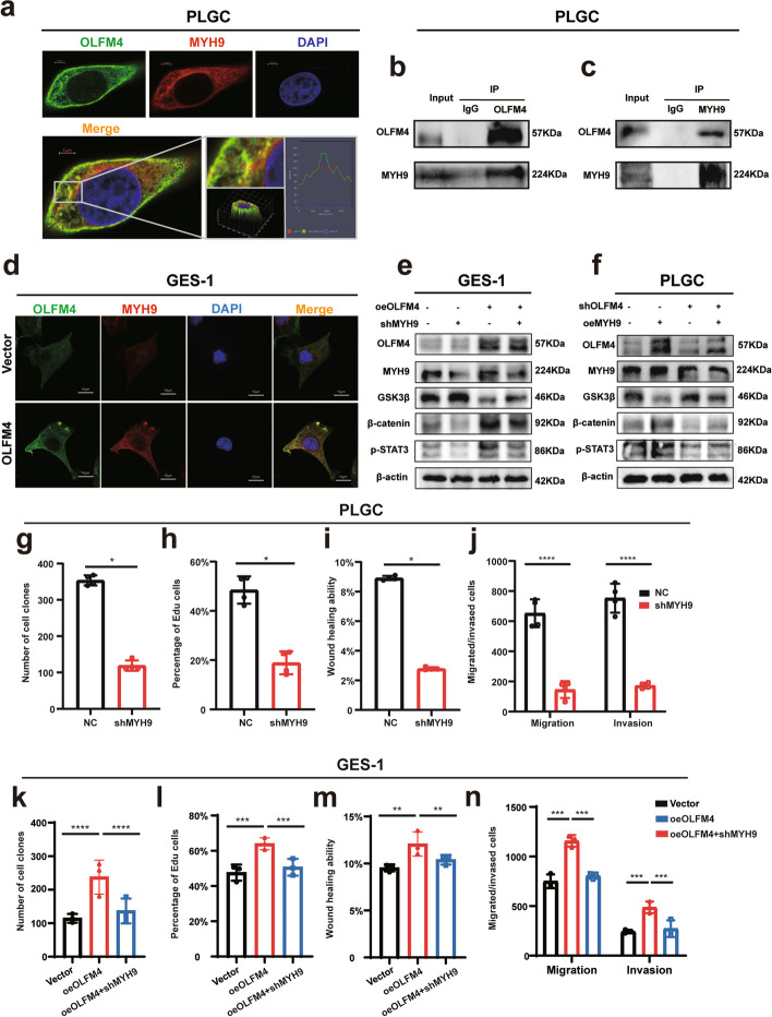
Fig. 5.
OLFM4 interaction with MYH9 stimulated activation of the Wnt pathway: a The figures demonstrated the co-localization of OLFM4 (green) and MYH9 (red) in fixed and immunostained cells. Cells were imaged using confocal microscopy, and the overlay image (yellow) indicated regions of co-localization, where both proteins were present in close proximity. Nuclei were stained with DAPI (blue) to provide cellular landmarks. The scale bar representeds 5 μm. b-c In the forward Co-IP, an antibody specific to OLFM4 was used to immunoprecipitate the protein complex from cell lysates. Western Blot analysis subsequently revealed the presence of MYH9 in the immunoprecipitated complex, indicating that MYH9 was binding with OLFM4. Conversely, in the reverse Co-IP, an antibody specific to MYH9 was employed to immunoprecipitate the associated proteins. Western Blot analysis confirmed the presence of OLFM4 in the immunoprecipitated complex. d After overexpressing OLFM4 in GES-1 cells, the expression level of MYH9 was evaluated through confocal immunofluorescence analysis. The scale bar representd 10 μm. e-f Western Blot analysis demonstrated the effects of overexpressing OLFM4 followed by knocking down MYH9, or overexpressing MYH9 followed by knocking down OLFM4 on the expression levels of Wnt pathway signals. g-n Statistical analysis of repeated experimental data for cell cloning, EdU, wound healing, and transwell assays after MYH9 knockdown in PLGC cells (shMYH9) or in oeOLFM4 cells. Statistics were expressed as mean ± SD. *p < 0.05, **p < 0.01, ***p < 0.001, ****p < 0.0001

