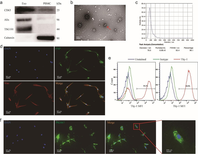
Fig. 2.
Plasma exosomes were taken up by human OFs. (a-c) Characterization of Pla-Exos. (a) Western blot analysis identified positive expression of exosomes markers CD63, Alix and TSG101, with the negative expression of Golgi marker Calnexin in the extraction samples. (b) Nano-Sight analysis indicated that the size of the extraction was approximately 100 nm. (c) The morphology of the extraction was examined via transmission electron microscope. (d, e) Characterization of OFs. (d) Immunofluorescence staining of OFs reveals the uniform expression of the fibroblast markers FSP and vimentin. Cells were stained with anti-FSP and anti-vimentin antibodies and Alexa Fluor 488/594-conjugated secondary antibodies (fluorescence microscopy, 20; scale bars, 50 μm). (e) OFs were analyzed for Thy-1 expression using flow cytometry (right). The Thy-1+ subset was obtained from parental OFs after 2 rounds of magnetic bead sorting. Thy-1+ OFs were > 99% positive for Thy-1 (left). (f) Pla-Exos (green) were transferred into OFs (blue) (fluorescence microscopy, 20). FSP fibroblast surface protein, VIM vimentin

