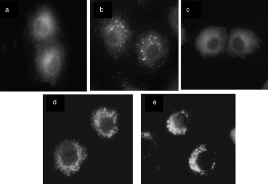
Figure 1.

Subcellular localization of hematoporphyrin monomethyl ether (HMME) in NuTu‐19 cells derived from adenocarcinoma of Fischer 344 rat. Fluorescence images of the cells double‐stained with HMME and mitochondria fluorescent probe (Rhodamine‐123; Sigma) or cytolysosome fluorescent probe (Lucifer Yellow; Sigma) are shown. Cells were observed at various time intervals (0.5, 1, 2, 3, 6, and 12 h) following coincubation of HMME and Rhodamine‐123 or Lucifer Yellow. (a) HMME + Rhodamine‐123; (b) HMME + Lucifer Yellow; (c) HMME alone; (d) Rhodamine‐123 alone; (e) Lucifer Yellow alone. All photos were taken 3 h after incubation.
