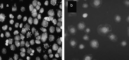
Figure 3.

Mitochondria damage in NuTu‐19 cells, derived from adenocarcinoma of Fischer 344 rat, following hematoporphyrin monomethyl ether photodynamic treatment. (a) Orange fluorescent spots in control cells indicate the intact mitochondria. (b) Mitochondria fluorescent spots disappeared completely and there were only diffused weaker green fluorescent spots in the cytoplasm, indicating the mitochondria damage.
