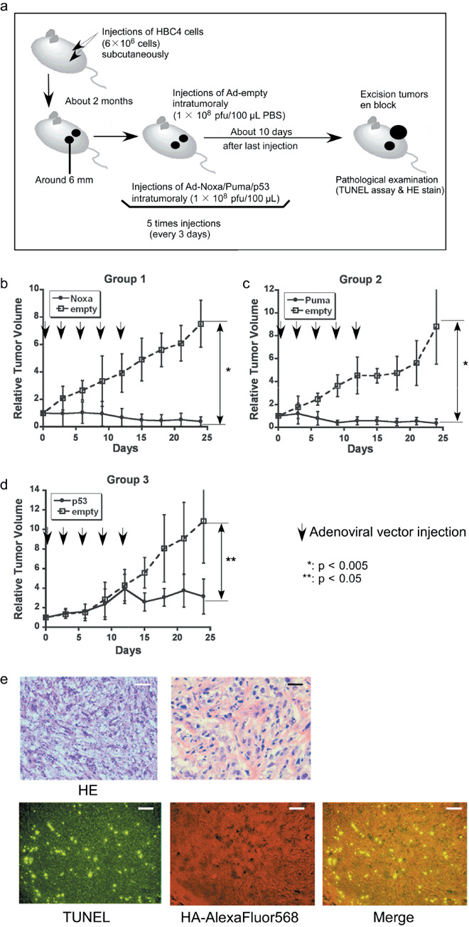
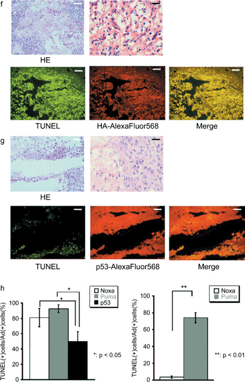
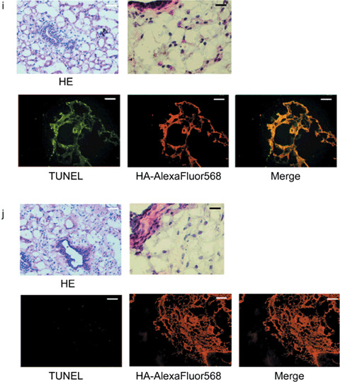
Figure 2.


Antitumor effect of adenoviral proapoptotic gene expression in vivo. (a) Assay procedures of adenoviral gene expression in a xenograft tumor model. Suppression of tumor growth by treatment with (b) Ad‐HA‐Noxa, (c) Ad‐HA‐Puma, and (d) Ad‐empty. At the indicated points, the major axis, minor axis, and height of tumors were measured. Then, tumor volume was estimated approximately as ellipsoid and normalized by the volume of the tumor just before adenoviral gene therapy. The experiments were carried out in quadruplicate and the results are presented as the mean ± SD. (e–g,i,j) Histopathological examinations of tumors and their peripheral tissues. These (e–g) tumors and (i,j) peripheral tissues were sliced and stained with hematoxylin–eosin (HE), TdT‐mediated dUTP‐biotin nick‐end labeling (TUNEL), and immunohistochemistry using anti‐HA‐antibody after adenoviral gene therapy with (e,j) Ad‐HA‐Noxa, (f,i) Ad‐HA‐Puma, and (g) Ad‐empty. White bars = 200 µm, black bars = 50 µm. (h) Apoptotic cell death in adenovirus‐infected tumors (left graph) and their peripheral tissues (right graph). The rates of TUNEL‐positive cells were measured in 10 independent focuses. The results are presented as the mean + SD.
