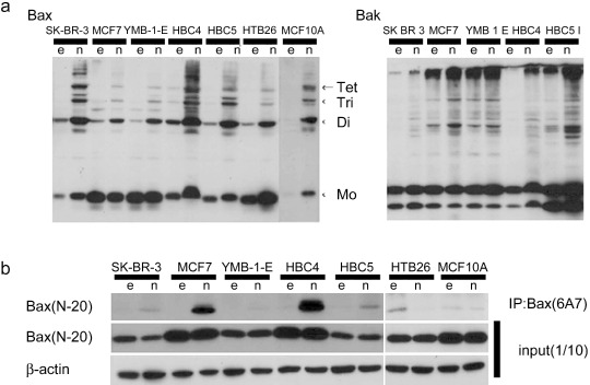
Figure 3.

Differences in the activation of Bax and Bak depend on cell lines. Tumor cells and MCF10A were infected with 5 multiplicity of infection of Ad‐empty (e) or Ad‐HA‐Noxa (n) for 12 h, and then whole‐cell lysates were collected. (a) Polymer formation of Bax or Bak protein. The mitochondrial fraction was collected from cell lysates, followed by cross‐linking between Bax molecules and 1,6‐bismaleimidohexane. Then, fractions were immunoblotted with anti‐Bax (left panel) or anti‐Bak (right panel) antibodies. (b) Immunoblot of activated Bax. Whole‐cell lysates were analyzed by immunoprecipitation (IP) with anti‐Bax (6A7) antibody, followed by immunoblotting with anti‐Bax (N‐20) antibody (upper panel). To verify that these cells expressed Bax protein, whole‐cell lysates were also immunoblotted using anti‐Bax (N‐20) and anti‐β‐actin antibodies (middle and lower panels).
