
Figure 1.
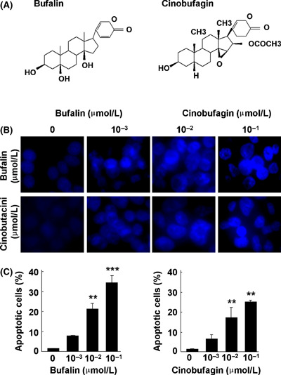
Bufalin and cinobufagin induce apoptosis in HepG2 cells. (A) Structure of bufalin and cinobufagin. (B) Representative microphotographs of Hoechst 33258 staining (original magnification, ×400). (C) Proportion of apoptotic cells. The data represent mean ± SD (n = 3). **P < 0.01 and ***P < 0.001 versus untreated controls.