
Figure 3.
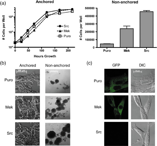
Sdpr protein expression in non‐transformed, mitogen‐activated protein kinase (MEK)‐transformed, and Src‐transformed cells. The genomic Sdpr gene in NIH 3T3 cells was tagged with green fluorescence protein (GFP) to produce an endogenously regulated fusion protein. These cells were then transfected with v‐Src, oncogenic MEK, or the empty parental expression vector (Puro) as controls. (a) Cells were plated (20 000 per well) on standard or low‐attachment tissue‐culture plates to monitor anchored or non‐anchored cell growth at the time points indicated. Only the Src‐ and MEK‐transfected cells were capable of anchorage‐independent cell growth. (b) Phase contrast image of anchored and non‐anchored cells at 3 days (bar = 200 microns) and 10 days (bar = 500 microns) growth, respectively. (c) GFP‐tagged Sdpr was observed in non‐transformed and transformed cells by confocal microscopy. Sdpr expression was reduced by transformation with Src, but not MEK (bar = 20 microns). DIC, differential interference contrast.