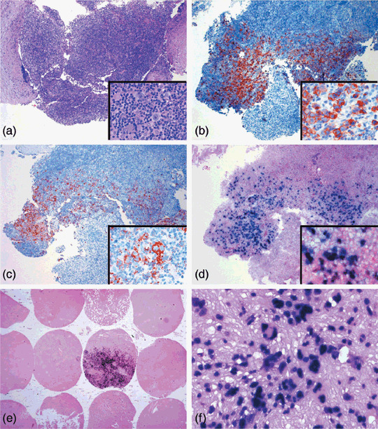
Figure 1.

Epstein–Barr virus (EBV)‐encoded early RNA (EBER1) in situ hybridization for Hodgkin lymphoma (HL) cases shows positive signals in tumor cells with larger nuclear appearance. (a–d) The tumor cells of this nodular sclerosis Hodgkin's lymphoma case best appreciated by (a) hematoxylin–eosin (HE) section are positive for (b) CD30 and (c) CD15. (d) The distribution of EBER‐positive tumor cells corresponds to that seen in the HE section. (e) Sections of tissue microarray for cases from Veterans General Hospital Taipei show positive signals mainly in large nucleated tumor cells, (f) although a few inflammatory cells show weaker staining. (a–d) Original magnification 40×, inset 400×; (e) original magnification 10×; (f) original magnification 200×.
