
Figure 4.
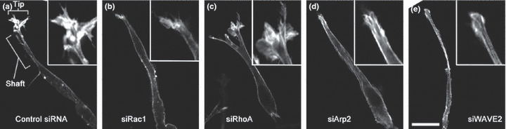
Pseudopodial F‐actin in cells showing elongated motility in 3D. (a) Control HT1080 cells form well‐developed tip and shaft structures. (b–e) HT1080 cells treated with Rac1, RhoA, actin‐related protein (Arp)2, and Wiskott–Aldrich syndrome protein family verprolin‐homologous protein (WAVE)2 siRNA, respectively. Note that the tip structures are lost in Rac1, Arp2, and WAVE2 siRNA treated cells. Cells are embedded in type I collagen gels. F‐actin is visualized by staining with fluorescently labeled phalloidin. Insets show magnification of the tip structure. Details are described in reference( 61 ). Bar, 20 μm.