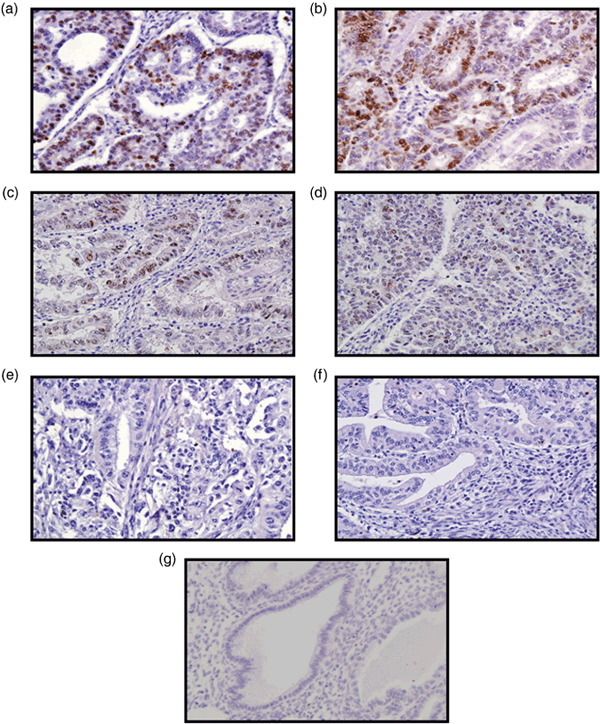
Figure 1.

Immunohistochemical analysis of Ret finger protein (RFP) in endometrial cancer. (a,b) Strongly positive, (c) moderately positive, (d) weakly positive, and (e) negative cases. (f) No primary antibody. (g) Normal endometrium. When greater than 10% of the cancer cells were stained with the RFP antibody regardless of staining intensity, the specimen was categorized as positive for RFP expression.
