Figure 1.

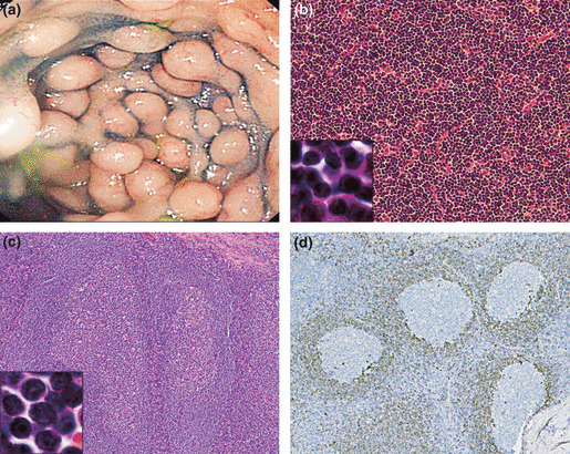
(a) Endoscopy of the colon of Case 11 showed multiple lymphomatoid polyposis involving the entire length of the colon. (b) Histological features of the small‐cell variant of mantle cell lymphoma (small‐MCL) in Case 5 showing a diffuse growth pattern without proliferation centers (original magnification ×200). At higher magnification (original magnification ×2500), small atypical lymphocytes with scant cytoplasm and dense nuclear chromatin are seen. Mitotic features were very rare (H&E). (c) The initial biopsy specimen of a tonsil obtained 4 years earlier from Case 12 shows pronounced follicular hyperplasia (original magnification ×100). The small‐MCL cells (original magnification ×2500) have round or oval small nuclei with dense nuclear chromatin and resemble mature B cells (H&E). (d) In the same specimen shown in (c), CCND1‐positive small lymphoid cells are thinly spread in the mantle zones and very few, if any, tumor cells have spread into interfollicular areas (original magnification ×100).
