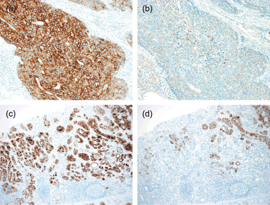
Figure 4.

Immunohistochemistry of α‐fetoprotein (AFP) in glypican 3 (GPC3)‐expressing gastric carcinomas (GC). Parallel immunostaining of (a,c) GPC3 and (b,d) AFP demonstrates both immunoreactive cells in the (a,b) hepatoid pattern and (c,d) clear‐cell tubular pattern of GPC3‐expressing GC. The numbers of cells immunoreactive for AFP were generally less than those of GPC3‐immunoreactive cells (×200).
