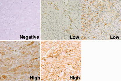
Figure 1.

Immunohistochemical grading of tumor specimens. Representative sections of osteosarcoma specimens stained with the anti‐HLA class I monoclonal antibody EMR8‐5 are shown (original magnification, ×200). ‘Negative’ indicates that less than 5% of tumor cells were stained positively. ‘Low’ indicates a positive tumor cell number from 5 to 50%. ‘High’ indicates a positive tumor cell number of over 50%. As seen typically in the low‐grade section, endothelial cells were stained positively with EMR8‐5, which was used as an internal positive control. The same immunohistochemical grading was used for sections stained with the antibody against anti‐β‐2 microglobulin.
