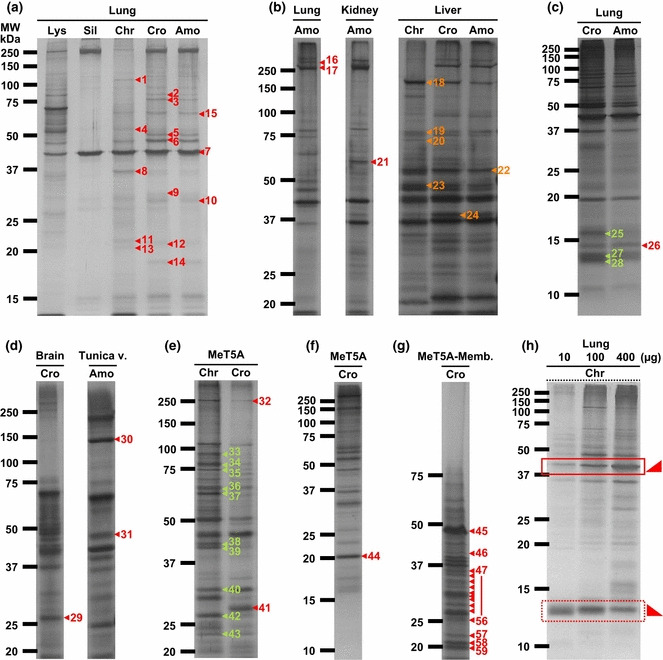
Figure 1.

Adsorption of specific proteins by asbestos fibers. Lysates from rat tissues or MeT5A mesothelial cells were incubated with each inorganic material and silver staining was performed after SDS‐PAGE. Asbestos exhibited higher adsorption than silica (Sil), and chrysotile (Chr) was the most adsorptive. (a–h) The panels show the original gels subjected to matrix‐assisted laser desorption ionization‐time of flight mass spectrometry (MALDI‐TOF/MS) analysis. The amount of rat lung lysate (Lys) loaded in panel (a) was 2 μg. Numbers correspond to the identified proteins listed in Table S1. The gels shown in each panel are different in the concentration of polyacrylamide and the combination of inorganic material and cell/tissue lysate. The origin of the rat lysate (e.g. lung, kidney, liver, etc.) used for the incubation is shown at the top and the species of the inorganic material is shown next to the top. (h) Chrysotile was incubated with 10, 100 or 400 μg of rat lung lysate and the proteins adsorbed onto chrysotile were analyzed by the coupling of SDS‐PAGE and silver staining. The square in a continuous line shows an increase in the amount of asbestos‐binding proteins as the total amount of proteins incubated increases. In contrast, the square in a dotted line shows the opposite. Cro, crocidolite; Amo, amosite; Tunica v., tunica vaginalis; MeT5A‐Memb., membrane fraction of MeT5A cells.
