
Figure 4.
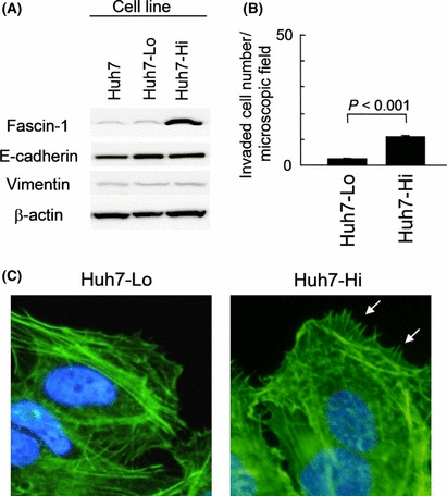
Characterization of Huh7‐derived cell lines, Huh7‐Hi and Huh7‐Lo, transfected with an expression construct of fascin‐1 cDNA. (A) Western blot analysis. (B) Transmembrane invasion assay (P‐value by Student’s t‐test). Error bar = SEM. (C) Fluorescently labeled phalloidin staining of actin filaments in Huh7‐Lo and Huh7‐Hi cells. More developed filopodia of Huh7‐Hi cells are indicated by white arrows.