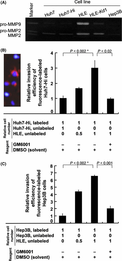
Figure 6.

Matrix metalloproteinases (MMPs) and coculture experiments. (A) Gelatin zymographic analysis of pro‐MMP2 and pro‐MMP9 in conditioned media. (B) Relative invasion efficiency of fluorescently labeled Huh7‐Hi cells cocultured with unlabeled Huh7‐Hi or HLE cells in the absence (−) or presence (+) of an MMP inhibitor, GM6001. The photograph shows orange fluorescence emitted from three invaded Huh7‐Hi cells labeled with CellTracker CM‐DiI reagent. The nuclei were stained with DAPI in blue. The three unlabeled cells were cocultured HLE cells. P‐values with an asterisk were obtained by the Jonckheere–Terpstra trend test; P‐values without an asterisk were obtained by Student’s t‐test. (C) Relative invasion efficiency of fluorescently labeled Hep3B cells cocultured with unlabeled Hep3B or HLE cells. Error bar = SEM.
