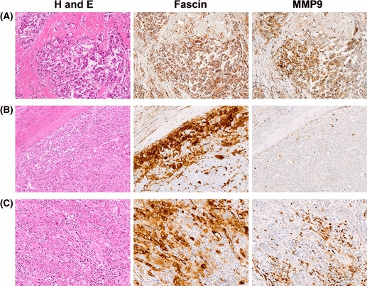
Figure 7.

Immunohistochemical analysis of MMP9 expression in fascin‐1‐positive primary hepatocellular carcinomas (HCCs). (A) Serial sections of a poorly differentiated HCC stained for H&E, fascin‐1 (fascin), or MMP9. Tumor cells at the invasive front are shown. They express both fascin‐1 and MMP9, and show an infiltrative growth pattern in the fibrous stroma. (B) Serial sections of a moderately differentiated HCC. Fascin‐1‐positive tumor cells with no detectable MMP9 expression are localized in the marginal area, and show a compressive growth pattern. (C) Serial sections of another moderately differentiated HCC. Although fascin‐1‐positive HCC cells produce no detectable MMP9, they show a highly infiltrative growth pattern accompanied by numerous MMP9‐positive inflammatory cells, most of which are neutrophils.
