
Figure 3.
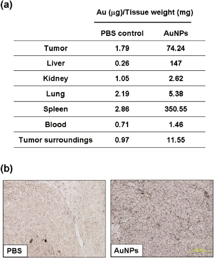
Biodistribution of gold nanoparticles (AuNP) in mice. (a) Twenty‐four hours after AuNP injection, tissues of tumor‐bearing mice were excised, processed, and used for AuNP detection using atomic absorption detection. (b) Silver staining of AuNP inside a tumor. Twenty‐four hours after AuNP injection, tumors were excised and paraffin‐embedded. Five‐micrometer thick sections from each representative specimen were obtained and then processed with the silver enhancement kit (bar = 200 µm). PBS, phosphate‐buffered saline.