Figure 3.

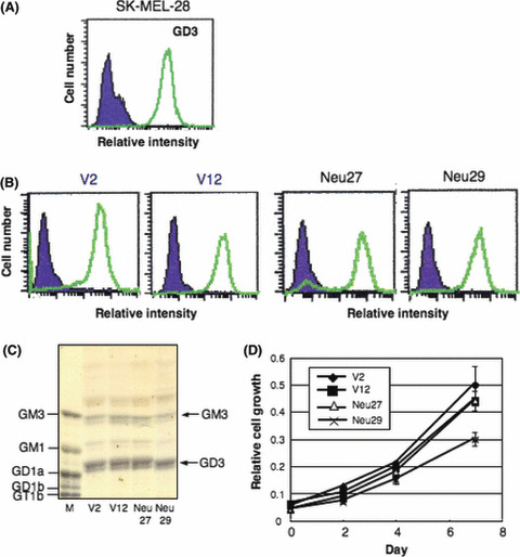
No changes in GD3 expression and cell growth in NEU3 transfectants. (A) Flow cytometry of GD3 expression in the parent line, SK‐MEL‐28. (B) GD3 expression in the transfected lines with vector alone and transfectant cell lines of NEU3 cDNA. Note expression of GD3 in Neu27 cells did not clearly change despite high NEU3 activity. (C) Thin‐layer chromatography patterns of gangliosides extracted from control lines and NEU3‐transfectant cell lines as detected by resorcinol spray. M, ganglioside marker (bovine brain gangliosides). (D) Cell proliferation of NEU3 transfectants. Cell growth of two each of controls and transfectant lines was compared using MTT assay.
