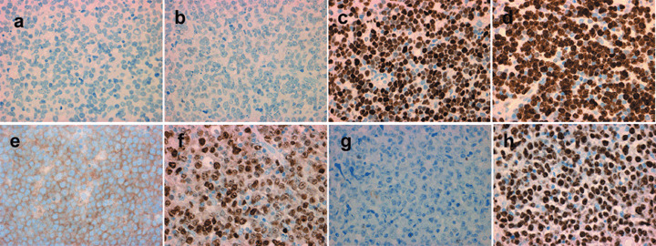
Figure 2.

The immunohistochemical panel for case 2 (upper row), belonging to the non‐germinal center B‐cell‐like group, with (a) CD10‐negative, (b) Bcl‐6‐negative, and (c) MUM1‐positive staining, and (d) high proliferative activity as labeled by Ki‐67. The immunohistochemical panel for case 6 (lower row), belonging to the germinal center B‐cell‐like group, with (e) CD10‐positive, (f) Bcl‐6‐positive, and (g) MUM1‐negative staining, and (h) high proliferative activity as labeled by Ki‐67.
