
Figure 4.
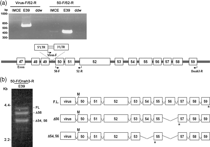
Fusion transcript between Dnah3 and retrovirus. (a) A fusion transcript identified using reverse transcription‐polymerase chain reaction (RT‐PCR). Left, RT‐PCR using retrovirus (virus‐F) and Dnah3 exon 52 (52‐R) primers. A fusion transcript is found in the E39 clones. In contrast, the wild‐type Dnah3 transcripts are detected both in IMCE and the E39 clone using Dnah3‐specific 50‐F and 52‐R primers. (b) Alternative transcripts observed in the E39 clone. RT‐PCR shows three different products. Structures of the retrovirus–Dnah3 chimeric transcripts are shown in the right panel. M, putative translation initiation; *, termination codons.