
Figure 3.
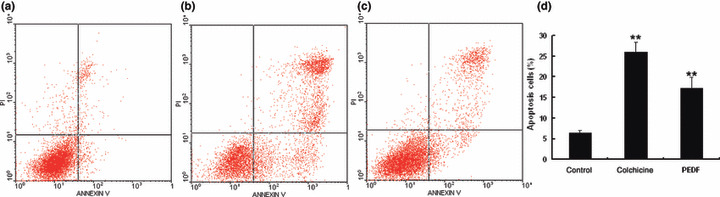
Quantitative analysis of endothelial cell apoptosis induced by pigment epithelium‐derived factor (PEDF). Human umbilical vein endothelial cells (HUVECs) were treated with PEDF for 24 h. Apoptotic cells were quantified by flow cytometry. (a) Negative control treated with PBS; (b) positive control treated with colchicine; (c) HUVECs treated with PEDF at 200 nmol/L; (d) statistical analysis of apoptosis results. Values significantly higher than controls (**P < 0.01) are indicated.