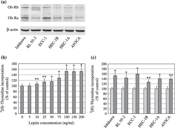
Figure 1.

Expression of the leptin receptor and effect of leptin on the proliferation of endometrial cancer cells. (a) Western blot analysis of leptin receptor expression, long form (Ob‐Rb, 120 kDa) and short form (Ob‐Ra, 90 kDa), in endometrial cancer cells. Equal loading and transfer were shown by repeat probing with β‐actin. (b) Serum‐starved Ishikawa cells were treated for 24 h with increasing concentrations of leptin and (c) six lines of serum‐starved endometrial cancer cells were treated with a 100 ng/mL dose of leptin for 24 h. DNA syntheses were determined by [3H] thymidine incorporation assay. *P < 0.01 and **P < 0.05 compared with untreated control cells, respectively.
