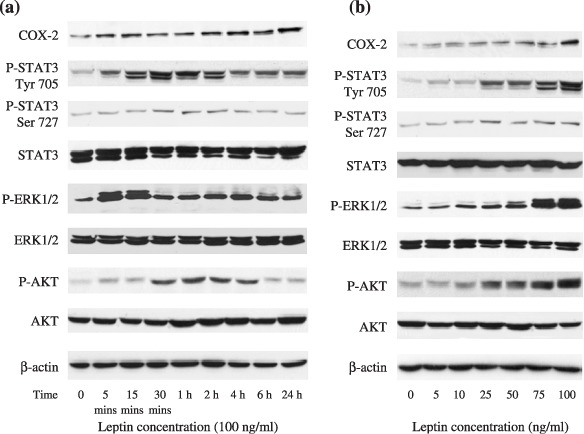
Figure 2.

Multiple intracellular pathways involved in the growth‐stimulatory effect of leptin on endometrial cancer cells. Serum‐starved Ishikawa cells were treated with leptin (100 ng/mL) for various intervals of time (a) and treated for 10 min with increasing concentrations of leptin (b). Protein expressions of cyclooxygenase (COX)‐2, total and phosphorylated forms of signal transducers and activators of transcription 3 (STAT3), extracellular signal‐regulated kinase (ERK1/2), as well as AKT were detected by western blot analysis. Equal loading and transfer were shown by repeat probing with β‐actin.
