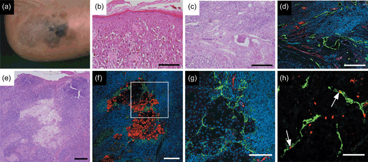
Figure 4.

Tumor and nodal lymphangiogenesis in cutaneous malignant melanoma. (a) Macroscopic appearance of early malignant melanoma in heel. Pigmented macules with irregular outline and remarkable variation in color illustrate superficial melanocyte spread during tumor progression. (b) Rounded melanocytes with atypical, hyperchromatic nuclei and abundant cytoplasm are scattered in a pagetoid pattern throughout epidermis. (c) Dermal invasion of melanoma cells develops tumor nests surrounded by tumor‐associated stroma during vertical growth in malignant progression. (d) Double immunofluorescence staining with anti‐von Willebrand factor antibody (red) shows enlarged blood vasculature. Podoplanin staining (green) shows prominent lymphatic vessel growth in tumor‐associated tissue. Tumor lymphangiogenesis can actively promote sentinel lymph node metastasis by tumorigenic melanoma cells. (e) Regional lymph nodes with metastatic cutaneous malignant melanoma (hematoxylin–eosin staining). (f) Double immunofluorescence staining using HMB‐45, a melanoma‐specific marker (red) in the corresponding area indicates metastatic melanoma cells that are closely associated with tumor‐associated podoplanin‐positive lymphatic vessels (green). (g) Vasculature in the corresponding field of Fig. 4f visualized using anti‐von Willebrand factor antibody for blood vessels (red), and D2‐40 for lymphatic vessels (green). Striking enlargement of tumor‐associated lymphatic vasculature might be associated with further distant spread of tumors. (h) Podoplanin‐positive lymphatic vessels (green) within metastatic lymph nodes stained with Ki‐67 (red) indicate active lymphatic proliferation (arrows). Scale bars, 100 µm (b,g); 200 µm (c–f); 50 µm (h). Nuclei are stained blue (diethylenetriaminepentaacetic acid).
