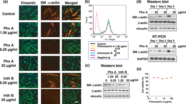
Figure 2.

Effects of phthoxazolin A (Phx A) and inthomycin B (Inth B) on myofibroblast differentiation. (a) Prostate stromal cells (PrSC) were cultured with the indicated concentrations of test compounds for 3 days. The presence of myofibroblasts was evaluated by immunofluorescence using anti‐vimentin (green) and anti‐smooth muscle (SM) α‐actin (red) antibodies. Photos are representative immunofluorescence results of three independent experiments with similar results. Scale bar = 50 µm. (b) PrSC were cultured with test compounds for 3 days and the expression of vimentin was analyzed by flow cytometry. (c) PrSC were cultured with Phx A and Inth B for 3 days. Cell lysates were prepared and used for western blotting. (d) PrSC were cultured with 25 µg/mL Phx A for the indicated times. Cell lysates and total RNA were prepared and used for western blotting and reverse transcription–polymerase chain reaction (RT‐PCR), respectively. (e) PrSC were precultured for 3 days and then further cultured with the Phx A for 3 days. Cell viability was assessed using trypan blue. Values represent means of duplicate determinations (SE < 10%). All data are representative of three independent experiments with similar results. GAPDH, glyceraldehyde 3‐phosphate dehydrogenase.
