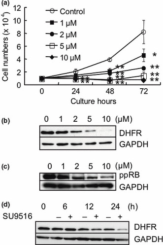
Figure 2.

The effects of SU9516 on growth, dihydrofolate reductase (DHFR), and retinoblastoma (RB) proteins in Jurkat cells. (a) Numbers of cells measured every 24 h were compared in the presence or absence of various concentrations of SU9516 by counting the cells a Trypan blue dye exclusion test. The data represent means of triplicate and the bars show SDs (*P < 0.05, **P < 0.01). (b) Whole‐cell extracts from Jurkat cells treated with the indicated concentrations of SU9516 or vehicle control (dimethyl sulfoxide, DMSO) for 24 h were subjected to a Western blot analysis using DHFR and GAPDH antibodies. (c) Whole‐cell extracts from Jurkat cells treated with the indicated concentrations of SU9516 or DMSO for 24 h were subjected to a Western blot analysis using phosphorylated pRB (Ser780) and GAPDH antibodies. (d) Whole‐cell extracts from Jurkat cells treated with DMSO or 2 μM of SU9516 for the periods indicated were subjected to a Western blot analysis using DHFR and GAPDH antibodies. GAPDH, glyceraldehyde 3‐phosphate dehydrogenase.
