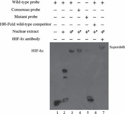
Figure 5.

Analysis of hypoxia‐inducible factor 1 (HIF‐1) binding to the hypoxia responsive element (HRE) motifs in the S100A4 gene in vitro using EMSA. BGC823 cells were exposed to normoxic or hypoxic mimicking conditions for 24 h prior to preparation of nuclear extracts. Various probes were incubated with nuclear extracts from CoCl2‐treated or nontreated BGC823 cells. Lane 1: labeled S100A4‐HREwt probe without nuclear extract; Lane 2: labeled S100A4‐HREwt probe incubated with nuclear extract from nontreated cells formed no specific complex; Lane 3: labeled S100A4‐HREwt incubated with the nuclear extracts from CoCl2‐treated cells forming a complex; Lane 4: labeled Epo‐HREwt as consensus oligonucleotide probe incubated with the nuclear extracts from CoCl2‐treated cells forming a complex; Lane 5: labeled S100A4‐HREmu probe did not form a complex; Lane 6: competition experiments performed with the S100A4‐HREwt probe using a 100‐fold molar excess of unlabeled S100A4‐HREwt oligonucleotides; Lane 7: supershift analysis of HIF‐1 bound to the S100A4‐HREwt probe performed using anti‐HIF‐1α antibody; *nuclear extracts from the CoCl2‐treated cells.
