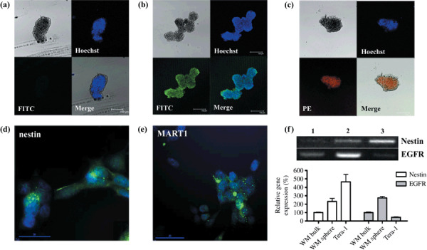
Figure 2.

Melanoma spheroid cells express nestin and epidermal growth factor receptor (EGFR). A whole sphere was fixed at 4% paraformaldehyde and stained with Hoechst dye solution to visualize nuclei with the UV light emission wavelength at 495 nm. (a–c) Immunocytochemistry with anti‐human melanoma antigen recognized by T‐cells (MART)1 (a), nestin (b), and EGFR (c) antibody. Spheroid cells did not show detectable MART1 signals. Overall nestin (FITC) and EGFR (PE) expressions in the spheroid cells are shown. Spheres approximately 150 µm in diameter with a central dark shadow were considered as the minimum size. (d–e) Immunocytochemistry for nestin and MART1 in the attached WM‐266‐4 cells. Attached WM‐266‐4 cells expressed nestin in some cells of cytosol with filamentous morphology. Representative of three independent experiments. (f) Semi‐quantitative RT‐PCR results for nestin and EGFR. Lane 1: attached WM‐266‐4 bulk cells. Lane 2: WM‐266‐4 spheroid cells. Lane 3: Teratocarcinoma cells (Tera‐1) cells as a stem cell–positive control.
