
Figure 2.
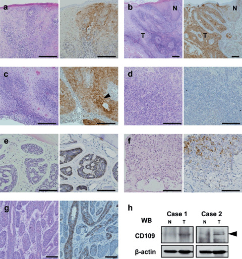
CD109 expression in oral tumor tissues. (a) Carcinoma in situ. (b) Well‐differentiated squamous cell carcinoma (SCC). (c) Well‐differentiated SCC with keratinizing regions (arrowhead). (d) Poorly differentiated SCC (negative case). (e) Adenoid cystic carcinoma. (f) Mucoepidermoid carcinoma. (g) Basal cell adenocarcinoma. Left panels, hematoxylin–eosin stain; right panels, CD109 immunostaining. Scale bars: a,b,c,d, 200 µm; e,f,g, 100 µm. (h) Western blotting for CD109 expression in SCC and adjacent normal tissues. Lysates were prepared from well‐differentiated SCC and adjacent normal tissues of two patients. N, normal tissue; T, tumor tissue.