
Figure 6.
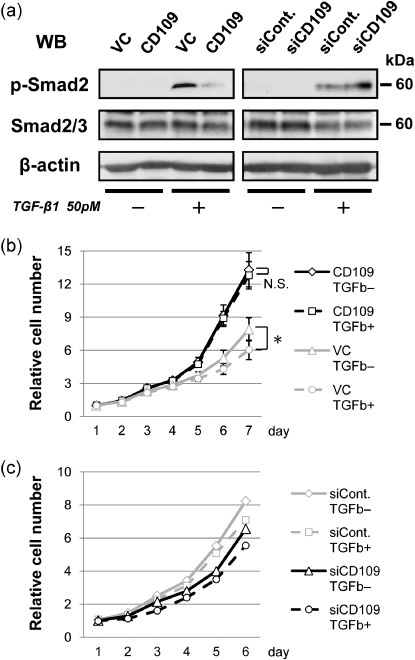
CD109 attenuates TGF‐β/Smad2 signaling. (a) Western blotting for assessment of Smad2 phosphorylation induced by TGF‐β1 (50 pM) in CD109‐overexpressing cells (left panel), and CD109‐knockdown cells (right panel). After 1 h stimulation with TGF‐β1, lysates of SAS‐VC and SAS‐CD109 cells, or SAS cells transfected with control and CD109 siRNA (siCont. and siCD109), were subjected to western blotting with antiphospho‐Smad2 (p‐Smad2), antitotal Smad2/3, and anti‐β‐actin antibodies. (b) Proliferation analyses of SAS‐VC and SAS‐CD109 cell lines in the presence or absence of TGF‐β1 (TGF‐β+ or TGF‐β–). The means ± SD are shown (bars). *P < 0.01, using the Student's t‐test. N.S., not significant. (c) Proliferation analyses of knockdown cells in the presence or absence of TGF‐β1.