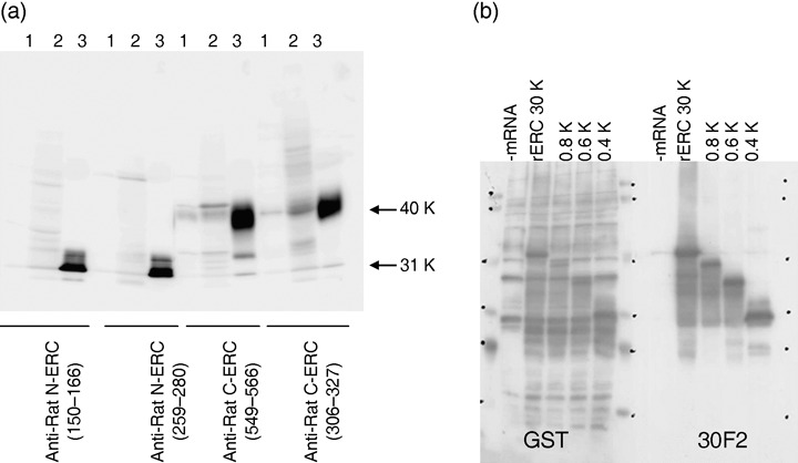
Figure 1.

Characterization of anti‐ERC/mesothelin antibodies. (a) Western blot analysis lane 1, MeET‐4 cell lysate; lane 2, lysates of MeET‐4 subcutaneously transplanted into nude mouse; and lane 3, culture supernatant of rat ERC/CHO transfectant. (b) The Epitope mapping of antirat ERC/mesothelin MoAb (30F2). Four different molecular sizes of the rat ERC/mesothelin fused with GST protein were produced and underwent Western blot analysis by 30F2 MoAb. 30F2MoAb reacted with all the GST‐fusion proteins.
