
Figure 4.
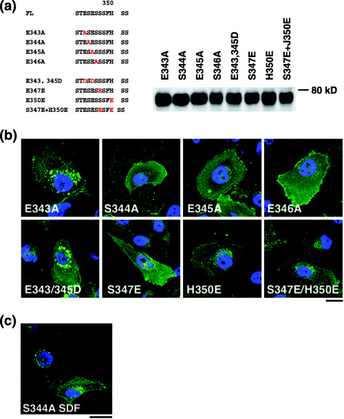
Characterization of the SDF‐1α‐independent internalization motif of CXCR4. (a) Left, amino acid sequences of CXCR4 FL and substitution mutants. Letters in red indicate introduced mutations. Right, protein expression of each mutant in 293T cells was verified by Western blot analysis. (b) Confocal micrographs of NP2 cells expressing each mutant. The blue signal represents the Hoechst‐stained nucleus. (Original magnification, ×630; bar, 10 µm.) (c) NP2 cells expressing the S344A mutant treated with SDF‐1α for 2 h are shown. The blue signal represents the Hoechst‐stained nucleus. (Original magnification, ×630; bar, 10 µm.)