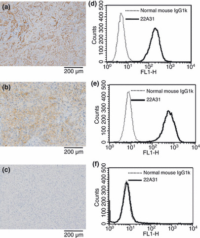
Figure 1.

Expression of C‐ERC/mesothelin in ACC‐MESO‐4, NCI‐H226, and Huh7 cells. Immunohistochemical localization of C‐ERC/mesothelin in ACC‐MESO‐4 cell‐ (a), NCI‐H226 cell‐ (b), and Huh7 cell‐ (c) derived xenografts. Indicated panel is representative of xenograft specimens. Original magnification, ×100. Surface expression of C‐ERC/mesothelin on ACC‐MESO‐4 cells (d), NCI‐H226 cells (e), and Huh7 cells (f) analyzed by flow cytometry. Solid line denotes staining of 22A31 and detected with Alexa Flour‐488. Dashed line denotes staining normal mouse IgG1κ.
