
Figure 3.
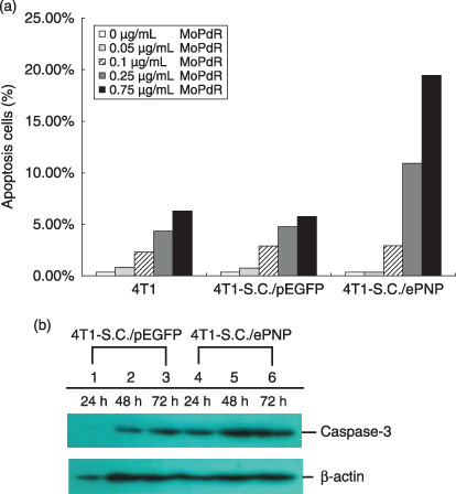
Assessment of apoptosis in 4T1 murine mammary tumor cells. (a) Enhancement of apoptosis. After incubation with 6‐methoxypurine 2′‐deoxyriboside (MoPdR) at different concentrations (0–0.75 µg/mL) for 96 h, harvested cells were fixed overnight with precooled ethanol and treated with 50 µg/mL propidium iodide (Sigma Chemical) and 10 µg/mL RNaseA for 30 min at 37°C. A minimum of 1.5 × 10⁴ events were analyzed on a fluorescence‐activated cell sorting flow cytometer with an argon laser tuned at 488 nm and use of CellQuest software (Becton Dickinson, Rutherford, NJ). Results are expressed as the percentage of apoptotic cells after S. typhimurium carrying pEGFP‐c1 (SC/pEGFP) and S. typhimurium carrying pEGFP‐c1‐ePNP (SC/ePNP) treatment with respect to the same cells non‐treated, in combination with MoPdR. (b) Western blot analysis. The infected cells were treated with 5 µg/mL MoPdR and total cellular proteins were isolated at indicated time points and subjected to Western blot analysis using anti‐caspase and anti‐β‐actin antibody.