
Figure 1.
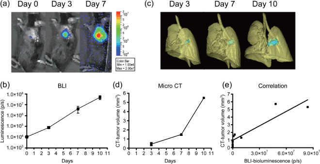
Non‐invasive monitoring of pulmonary tumor growth in an orthotopic lung cancer implantation model. Mice were intrapulmonary inoculated with luciferase‐expressing mouse Lewis Lung Carcinoma cell line (3LL‐luc2) cells (1000 cells in 20 µL volume) on day 0, and then monitored for orthotopic tumor growth by bioluminescence imaging (BLI) (a,b) or microcomputed tomography (micro‐CT) (c,d) on indicated days after tumor inoculation. Images of the 3D‐reconstituted tumor colony are shown in blue. Data represent mean ± SEM. n = 3, except CT images on day 10 (n = 2). (e) Relationship between BLI of tumor and tumor volume reconstituted from micro‐CT images.