
Figure 2.
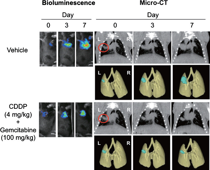
Bioluminescence imaging (BLI) and microcomputed tomography (micro‐CT) images of orthotopically implanted pulmonary tumor growth treated with combination chemotherapy. Mice were intrapulmonary inoculated with luciferase‐expressing mouse Lewis Lung Carcinoma cell line (3LL‐luc2) cells (1000 cells in 20 µL volume) on day 0, and then monitored for orthotopic tumor growth by BLI and micro‐CT. Images of the 3D‐reconstituted tumor colony are shown in blue. Mice were randomly assigned into study groups 7 days after the tumor inoculation and were treated with CDDP (cisplatin)/Gemcitabine. BLI and micro‐CT images were taken on indicated days after tumor inoculation. Representative images of five mice for each group are shown.