
Figure 2.
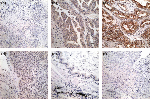
Immunohistochemical staining of SCC‐S2 in lung cancer tissue sections. (a) Negative staining in lung squamous cell carcinoma (−). (b) Positive immunostaining in lung adenocarcinoma (+), SCC‐S2 was localized mainly in the cytoplasm of cancer cells. (c) Strong SCC‐S2 staining in adenocarcinoma (++). (d) Weak SCC‐S2 immunostaining in lung squamous cell carcinoma. (e) SCC‐S2 was negative or was detected weakly in adjacent normal bronchial epithelia. (f) Negative controls were prepared by non‐immune rabbit IgG at the same dilution as for the primary antibody in tumor sample.