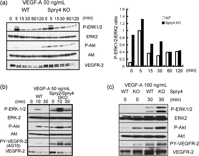
Figure 5.

Influence of Sprouty4 deficiency on vascular endothelial growth factor (VEGF)‐A signaling. (a,b) Wild‐type (WT) and Sprouty4 knockout (KO) mouse embryonic fibroblasts (MEFs) (a) or WT and Sprouty2/Sprouty4 double KO (DKO) MEFs (b), which stably expressed VEGFR‐2, were stimulated with 50 ng/mL VEGF‐A. The relative ratio of the band intensity of phosphorylated ERK1/2 versus that of total ERK2 is shown in the right (a). (c) Inferior venae cavae of WT and Sprouty4 KO were stimulated with 100 ng/mL VEGF‐A. (a–c) Cell extracts were immunoblotted with the indicated antibodies.
