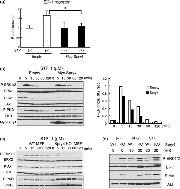
Figure 6.

Negative regulation of sphingosine‐1‐phosphate (S1P) signaling by Sprouty4. (a) Flag‐tagged control or Sprouty plasmid (0.5 µg) was transfected into mouse embryonic fibroblasts (MEFs) with the Elk‐1 reporter. Cells were treated with (+) or without (–) 1 µM S1P for 6 h and then analyzed with the luciferase assay. The results are expressed as fold‐increase in luciferase activity with S1P relative to that without S1P. Data shown are means ± SEM. *P < 0.05. (b) MEFs stably expressing empty or Myc‐tagged Sprouty4 plasmid were stimulated with 1 µM S1P. The relative ratio of the band intensity of phosphorylated ERK1/2 versus that of total ERK2 is shown in the right. (c) Wild‐type (WT) and Sprouty4 knockout (KO) MEFs were stimulated with 1 µM S1P. (d) Inferior venae cavae of WT and Sprouty4 KO were stimulated with 50 ng/mL basic fibroblast growth factor (bFGF) or 1 µM S1P. (b–d) Cell extracts were immunoblotted with the indicated antibodies.
