
Figure 2.
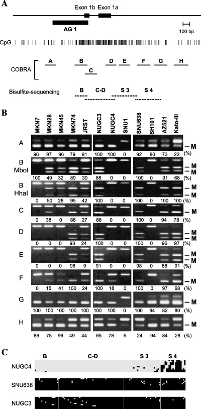
Methylation analysis of DFNA5 in gastric cancer cell lines. (A) Schematic representation of the DFNA5 CpG island. The solid box indicates the location of exon 1. The position of the DNA fragments obtained by methylated CpG island (CGI) amplification coupled with representational difference analysis is shown by the solid box designated as AG1. The solid horizontal lines indicate the regions analyzed by combined bisulfite restriction analysis (COBRA), the dotted lines the regions analyzed using bisulfite sequencing. (B) COBRA was carried out using eight sets of primers that covered the entire DFNA5 CGI. M, methylated alleles. Percentages of methylated alleles are shown below the column. Cell lines are shown on the top. (C) Bisulfite sequence analysis of DFNA5: □, unmethylated alleles; ▪, methylated alleles. Cell lines examined are shown on the left. The regions analyzed are shown on the top.