
Figure 4.
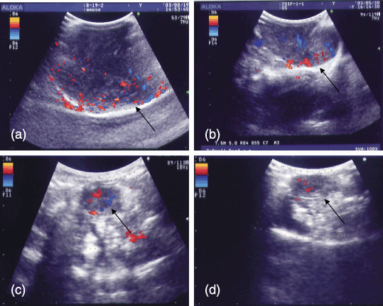
(a–d) Visualization of the intratumoral blood flow using non‐invasive contrasted color Doppler ultrasound (US) in FU‐MMT‐1 human uterine sarcoma growing subcutaneous on the backs of BALB/cA Jcl‐nu mice. Representative Doppler ultrasound pictures are taken at maximal signal intensity after injecting the US contrast agent (10 s after injection). The tumor vessel density in (c) the group of TNP‐470 treatment alone was lower than that of (a) the control (unpaired t‐test, P = 0.0007), whereas no significant difference was observed between (b) the treatment of US alone and the control (unpaired t‐test, P = 0.11). The reduction of tumor vessel density was significantly enhanced by (d) the combined treatment, in comparison to that of the US treatment alone (P = 0.00002), or the TNP‐470 alone (unpaired t‐test, P = 0.00009).