
Figure 6.
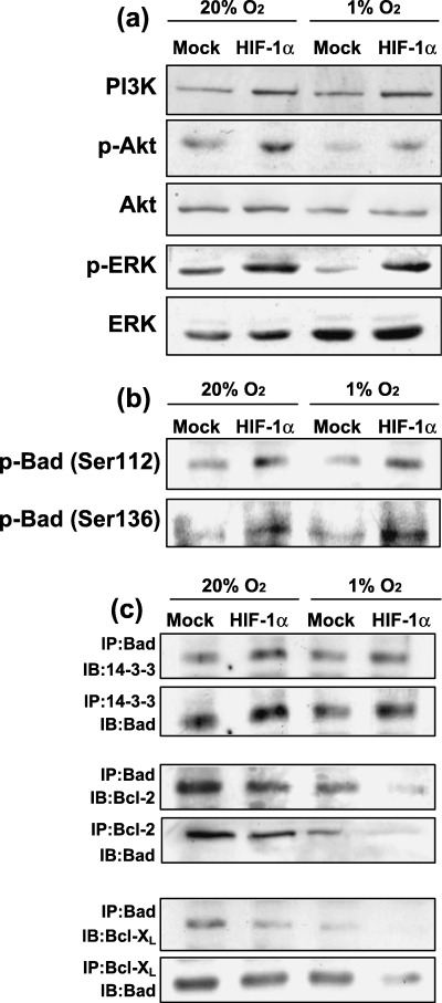
Effects of forced expression of HIF‐1α on the expression levels of PI3K, Akt and ERK and their phosphorylation levels. (a) Both cell types were cultured under normoxic and hypoxic conditions for 24 h. Cell lysates were then prepared and western blot analysis was performed to determine the levels of PI3K, Akt, p‐Akt (Ser473), total ERK1/2 and p‐ERK1/2. (b) Phosphorylation of Bad was determined by western blotting using antiphosphorylated Bad (Ser112 and Ser136) antibodies. (c) Hetero‐oligomerization between Bad, 14‐3‐3, Bcl‐2 and Bcl‐XL. Co‐immunoprecipitates were obtained using antibodies against each Bcl‐2 family protein and the obtained immunoprecipitates were blotted using each indicated antibody. The experiment was repeated three times and this is a representative example.