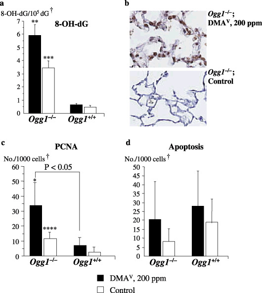
Figure 3.

Formation of 8‐hydroxy‐2′‐deoxyguanosine (8‐OH‐dG) and cellular proliferation and apoptosis indices in the lungs of Ogg1 −/– and Ogg1 +/+ mice treated with dimethylarsinic acid (DMAV) at 200 p.p.m. for 72 weeks. (a) 8‐OH‐dG (c) proliferating cell nuclear antigen (PCNA) and (d) apoptosis indices. (b) 8‐OH‐dG staining pattern in DMAV‐treated and control Ogg1 −/– mouse. †Normal‐appearing area; *Significant at P < 0.05 versus Ogg1 −/– control group; **Significant at P < 0.001 versus Ogg1 −/– control group; ***Significant at P < 0.0001 and ****Significant at P < 0.05 versus Ogg1 +/+ control group.
